Interrelationships between the Retinal Neuroglia and Vasculature in Diabetes
- PMID: 25003068
- PMCID: PMC4083021
- DOI: 10.4093/dmj.2014.38.3.163
Interrelationships between the Retinal Neuroglia and Vasculature in Diabetes
Abstract
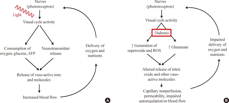
For years, diabetic retinopathy has been defined based on vascular lesions, and neural abnormalities were not regarded as important. This review summarizes evidence that the neural retina has important effects on the retinal vasculature under normal conditions, and the interaction between the retinal neuroglial cells and vascular function is altered in diabetes. Importantly, new evidence raises a possibility that abnormalities within retinal neuroglial cells (notably photoreceptors) might actually be causing or initiating the vascular disease in diabetic retinopathy.
Keywords: Diabetes mellitus; Diabetic retinopathy; Neurovascular coupling; Photoreceptors; Retina.
Conflict of interest statement
This work was funded by PHS grants EY00300 and EY022938, and by a Merit grant from the Veteran's Administration.
Figures
References
-
- Buerk DG, Riva CE, Cranstoun SD. Frequency and luminance-dependent blood flow and K+ ion changes during flicker stimuli in cat optic nerve head. Invest Ophthalmol Vis Sci. 1995;36:2216–2227. - PubMed
-
- Falsini B, Riva CE, Logean E. Flicker-evoked changes in human optic nerve blood flow: relationship with retinal neural activity. Invest Ophthalmol Vis Sci. 2002;43:2309–2316. - PubMed
-
- Logean E, Falsini B, Riva CE. Effect of chromatic flicker on circulation of the optic nerve. Klin Monbl Augenheilkd. 2001;218:345–347. - PubMed
-
- Riva CE, Logean E, Falsini B. Visually evoked hemodynamical response and assessment of neurovascular coupling in the optic nerve and retina. Prog Retin Eye Res. 2005;24:183–215. - PubMed
-
- Hardarson SH, Basit S, Jonsdottir TE, Eysteinsson T, Halldorsson GH, Karlsson RA, Beach JM, Benediktsson JA, Stefansson E. Oxygen saturation in human retinal vessels is higher in dark than in light. Invest Ophthalmol Vis Sci. 2009;50:2308–2311. - PubMed
Publication types
Grants and funding
LinkOut - more resources
Full Text Sources
Other Literature Sources