

Efficacy of Annona squamosa L in the synthesis of glycosaminoglycans and collagen during wound repair in streptozotocin induced diabetic rats
- PMID: 25003104
- PMCID: PMC4070582
- DOI: 10.1155/2014/124352
Efficacy of Annona squamosa L in the synthesis of glycosaminoglycans and collagen during wound repair in streptozotocin induced diabetic rats
Abstract
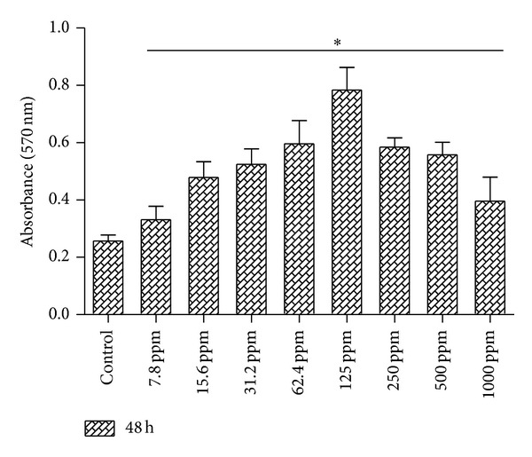
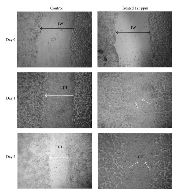
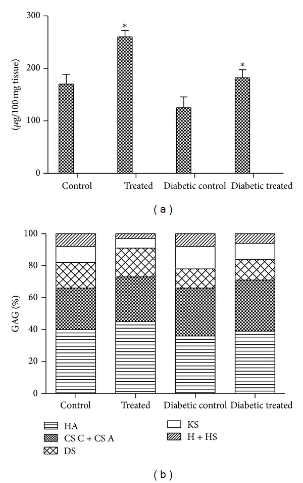
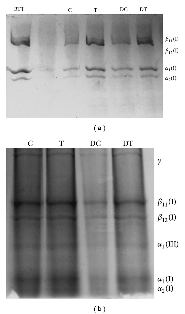
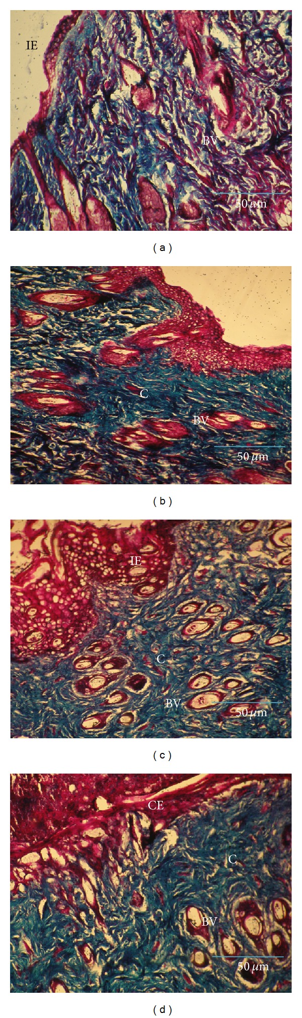
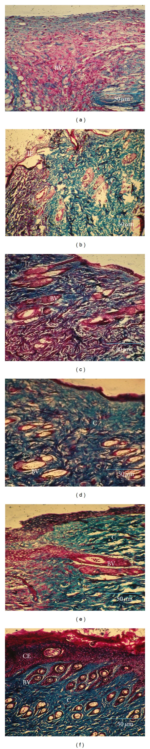
The aim of this work was to find out the effects of Annona squamosa on the formation of glycosaminoglycans and collagen during wound healing in normal and diabetic rats. Diabetes induced rats were segregated into 4 groups, each containing six animals. Groups I and III served as the normal and diabetic control while groups II and IV served as normal and diabetic treated. The animals were treated with 200 μL of Annona squamosa extract topically. The granulation tissues formed were removed on the 8th day and the amount of glycosaminoglycans (GAGs) and collagen formed was evaluated by sequential extraction and SDSPAGE, respectively. Histological evaluation was also carried out using Masson's trichrome stain. In vitro wound healing efficacy of A. squamosa in human dermal fibroblast culture (HDF) was also carried out. The fibroblasts treated with varying concentrations of A. squamosa were examined for proliferation and closure of the wound area and photographed. A. squamosa increased cellular proliferation in HDF culture. The granulation tissues of treated wounds showed increased levels of glycosaminoglycans (P < 0.05) and collagen which were also confirmed by histopathology. The results strongly substantiate the beneficial effects of A. squamosa on the formation of glycosaminoglycans and collagen during wound healing.
Figures
Similar articles
-
Efficacy of Annona squamosa on wound healing in streptozotocin-induced diabetic rats.Int Wound J. 2012 Dec;9(6):613-23. doi: 10.1111/j.1742-481X.2011.00924.x. Epub 2012 Jan 11. Int Wound J. 2012. PMID: 22233431 Free PMC article.
-
Antidiabetic and antioxidant activity of Annona squamosa extract in streptozotocin-induced diabetic rats.Singapore Med J. 2006 Aug;47(8):670-5. Singapore Med J. 2006. PMID: 16865205
-
Beneficial effects of Annona squamosa extract in streptozotocin-induced diabetic rats.Singapore Med J. 2008 Oct;49(10):800-4. Singapore Med J. 2008. PMID: 18946614
-
Antidiabetic activity of aqueous leaf extract of Annona squamosa in streptozotocin-nicotinamide type 2 diabetic rats.J Ethnopharmacol. 2004 Mar;91(1):171-5. doi: 10.1016/j.jep.2003.12.017. J Ethnopharmacol. 2004. PMID: 15036485
-
Diabetic Wound Healing: Factors, Mechanisms, and Treatment Strategies Using Herbal Components.Curr Drug Targets. 2025;26(6):367-381. doi: 10.2174/0113894501354898241220075327. Curr Drug Targets. 2025. PMID: 39791148 Review.
Cited by
-
Limited Treatment Options for Diabetic Wounds: Barriers to Clinical Translation Despite Therapeutic Success in Murine Models.Adv Wound Care (New Rochelle). 2021 Aug;10(8):436-460. doi: 10.1089/wound.2020.1254. Epub 2020 Dec 18. Adv Wound Care (New Rochelle). 2021. PMID: 33050829 Free PMC article.
-
Topical Application of Premna integrifolia Linn on Skin Wound Injury in Rats Accelerates the Wound Healing Process: Evidence from In Vitro and In Vivo Experimental Models.Evid Based Complement Alternat Med. 2022 Apr 13;2022:6449550. doi: 10.1155/2022/6449550. eCollection 2022. Evid Based Complement Alternat Med. 2022. PMID: 35463068 Free PMC article.
-
Glycosaminoglycan remodeling during diabetes and the role of dietary factors in their modulation.World J Diabetes. 2016 Feb 25;7(4):67-73. doi: 10.4239/wjd.v7.i4.67. World J Diabetes. 2016. PMID: 26962410 Free PMC article. Review.
-
Phenolic Compounds and In Vitro Antibacterial and Antioxidant Activities of Three Tropic Fruits: Persimmon, Guava, and Sweetsop.Biomed Res Int. 2016;2016:4287461. doi: 10.1155/2016/4287461. Epub 2016 Aug 25. Biomed Res Int. 2016. PMID: 27648444 Free PMC article.
-
A Randomized, Active Comparator-controlled Clinical Trial of a Topical Botanical Cream for Skin Hydration, Elasticity, Firmness, and Cellulite.J Clin Aesthet Dermatol. 2018 Aug;11(8):51-57. Epub 2018 Aug 1. J Clin Aesthet Dermatol. 2018. PMID: 30214668 Free PMC article.
References
-
- Falanga V. Wound healing and its impairment in the diabetic foot. The Lancet. 2005;366(9498):1736–1743. - PubMed
-
- Stadelmann WK, Digenis AG, Tobin GR. Physiology and healing dynamics of chronic cutaneous wounds. The American Journal of Surgery. 1998;176(2) 1:26S–38S. - PubMed
-
- Kumar B, Vijayakumar M, Govindarajan R, Pushpangadan P. Ethnopharmacological approaches to wound healing-exploring medicinal plants of India. Journal of Ethnopharmacology. 2007;114(2):103–113. - PubMed
-
- Iba Y, Shibata A, Kato M, Masukawa T. Possible involvement of mast cells in collagen remodeling in the late phase of cutaneous wound healing in mice. International Immunopharmacology. 2004;4(14):1873–1880. - PubMed
-
- Wysocki AB. Wound fluids and the pathogenesis of chronic wounds. Journal of Wound, Ostomy and Continence Nursing. 1996;23(6):283–290. - PubMed
Publication types
MeSH terms
Substances
LinkOut - more resources
Full Text Sources
Other Literature Sources
