. 2014 Jul 8;9(7):e101748.
doi: 10.1371/journal.pone.0101748. eCollection 2014.

β-Catenin inactivation is a pre-requisite for chick retina regeneration

Affiliations

β-Catenin inactivation is a pre-requisite for chick retina regeneration

Jie Zhu et al. PLoS One. .

Abstract

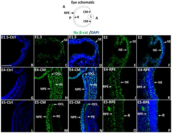
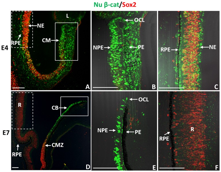
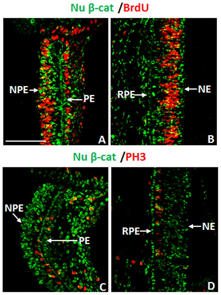
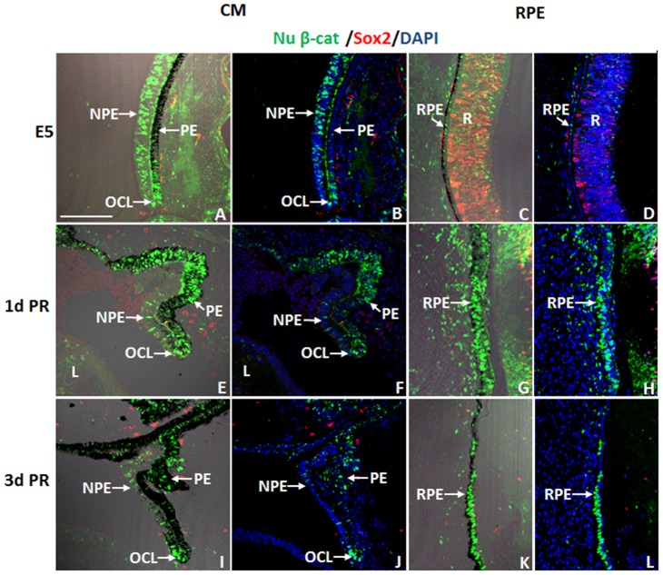
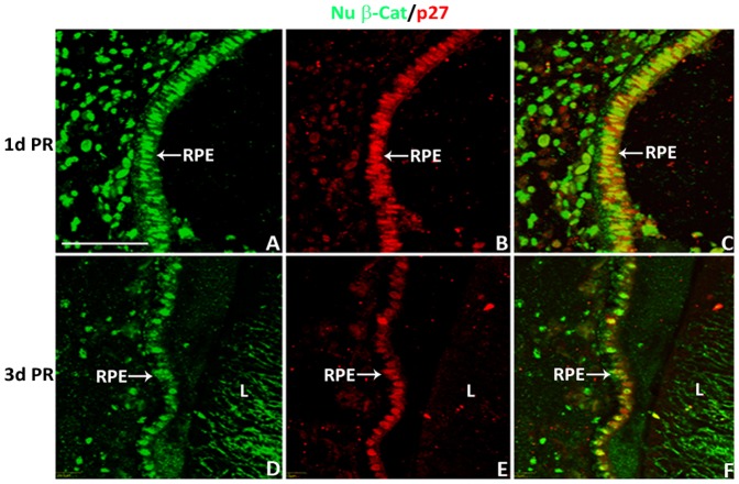
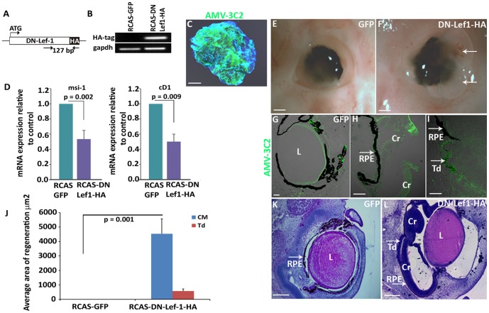
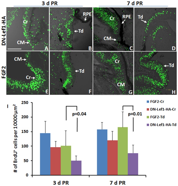
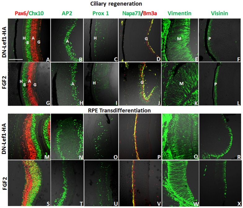
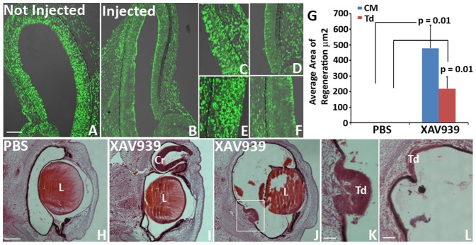
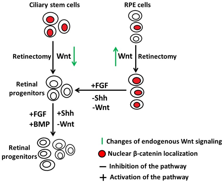
In the present study we explored the role of β-catenin in mediating chick retina regeneration. The chick can regenerate its retina by activating stem/progenitor cells present in the ciliary margin (CM) of the eye or via transdifferentiation of the retinal pigmented epithelium (RPE). Both modes require fibroblast growth factor 2 (FGF2). We observed, by immunohistochemistry, dynamic changes of nuclear β-catenin in the CM and RPE after injury (retinectomy). β-Catenin nuclear accumulation was transiently lost in cells of the CM in response to injury alone, while the loss of nuclear β-catenin was maintained as long as FGF2 was present. However, nuclear β-catenin positive cells remained in the RPE in response to injury and were BrdU-/p27+, suggesting that nuclear β-catenin prevents those cells from entering the cell cycle. If FGF2 is present, the RPE undergoes dedifferentiation and proliferation concomitant with loss of nuclear β-catenin. Moreover, retinectomy followed by disruption of active β-catenin by using a signaling inhibitor (XAV939) or over-expressing a dominant negative form of Lef-1 induces regeneration from both the CM and RPE in the absence of FGF2. Our results imply that β-catenin protects cells of the CM and RPE from entering the cell cycle in the developing eye, and specifically for the RPE during injury. Thus inactivation of β-catenin is a pre-requisite for chick retina regeneration.


Conflict of interest statement

Competing Interests: The authors have declared that no competing interests exist.

Figures

Figure 1
Figure 1. Nuclear β-catenin presence during chick eye development.
(A) Schematic diagram of the eye showing the anterior (A) and posterior (P) region of the eye as well as the location of the ciliary margin (CM), retina (R), retinal pigmented epithelium (RPE) and Lens (L). The orientation of the eye in the diagram applies to all images in this paper. (B–F) Immunohistochemistry showing the location of nuclear β-catenin (Nu β-cat) in the developing optic vesicles at E1.5 (HH 10) (B–D) and E2 (HH 13) (E–F). (G–P) Nu β-cat presence in the developing CM and RPE at E4 (HH 22–24) (G–K) and E5 (HH 27–28) (L–P). DAPI stains the nuclei of the cells in (B, D, F, G, I, K, L, N, P). B, G and L are the corresponding negative controls for E 1.5 (B), E4 (G) and E5 (L). SE: surface Ectoderm; NE: neuroepithelium; NPE: non-pigmented epithelium; PE: pigmented epithelium. OCL: optic cup lip. The scale bar in (B) represents 100 µm and applies to all panels.
Figure 2
Figure 2. Nuclear β-catenin+ cells in the CM and RPE of chick eyes do not co-express Sox2.
(A) Immunohistochemistry shows patterns of nuclear β-catenin (Nu β-cat)/Sox2 in E4 eyes. (B) Close-up image of the CM (boxed solid line) area in (A). (C) Close-up image of the RPE (boxed dash line) area in (A). (D) Patterns of Nu β-cat/Sox2 in E7 eyes. (E) Close-up image of the boxed (solid line) area in (D). (F) Close-up image of the boxed (dash line) area in (D). CM: ciliary margin; NPE: non-pigmented epithelium; PE: pigmented epithelium; OCL: optic cup lip; CB: ciliary body; CMZ: ciliary marginal zone; RPE: retinal pigmented epithelium; NE: neuroepithelium; R: retina. The scale bars represent 100 µm in all panels.
Figure 3
Figure 3. Many nuclear β-catenin+ cells in the CM and RPE of E4 chick eyes are not proliferating.
(A–B) The presence of nuclear β-catenin (Nu β-cat) and BrdU incorporation in the CM (A) and RPE (B) at E4. (C–D) Double immunohistochemistry for Nu β-cat and PH3 in the CM (C) and RPE (D) at E4. NPE: non-pigmented epithelium; PE: pigmented epithelium; RPE: retinal pigmented epithelium; NE: neuroepithelium. The scale bar in (A) represents 100 µm and applies to all panels.
Figure 4
Figure 4. Dynamic changes of nuclear β-catenin in the chick eye after injury.
(A–L) Presence of nuclear β-catenin (Nu β-cat) and Sox2 in the CM (A, B, E, F, I, J) and RPE (C, D, G, H, K, L) at E5 (A–D), at 1 d PR (E–H) and at 3 d PR (I–L). Panels A, C, E, G, I, K include DIC overlay and are equivalent to B, D, F, H, J, and L respectively. NPE: non-pigmented epithelium; PE: pigmented epithelium; OCL: optic cup lip; RPE: retinal pigmented epithelium; R: retina. DAPI stains the nuclei of the cells in B, D, F, H, J and L. Scale bar in (A) represents 100 µm and applies to all panels.
Figure 5
Figure 5. CM and RPE cells show low to no proliferative activity after retinectomy.
(A–H) Presence of nuclear β-catenin (Nu β-cat) and BrdU incorporation in the CM (A–D) and RPE (E–H) at 1 d PR (A, B, E, F) and 3 d PR (C, D, G, H). Panels A, C, E, G contain DIC overlay and are equivalent to B, D, F, and H respectively. NPE: non-pigmented epithelium; PE: pigmented epithelium; RPE: retinal pigmented epithelium; L: lens. DAPI stains the nuclei of cells in B, D, F and H. Scale bar in (A) represents 100 µm and applies to all panels.
Figure 6
Figure 6. Nuclear β-catenin+ RPE cells co-localize with cell cycle inhibitor p27 at 1 and 3 d PR.
(A–F) Presence of nuclear β-catenin (Nu β-cat) and p27 at 1 d PR (A–C) and 3 d PR (D–F). RPE: retinal pigmented epithelium; L: lens. Scale bar in (A) represents 50 µm and applies to all panels.
Figure 7
Figure 7. Nuclear β-catenin is absent in the CM and RPE during FGF2-induced regeneration.
Co-expression of nuclear β-catenin (Nu β-cat) and Sox2 in the CM (A–F) and RPE (G–L) at 1 d PR (A–C and G–I) and 3 d PR (D–F and J–L); (B,C) show the boxed area in (A); (E, F) show the boxed area in (D); (H, I) show the boxed area in (G); and (K, L) show the boxed area in (J). Panels A, D, G, and J have DIC overlay. Panels B, E, H, and K include DIC overlay and are equivalent to C, F, I, and L respectively. b = FGF2 bead; Cr = ciliary regeneration; Td = transdifferentiation; PE: pigmented epithelium; NPE: non-pigmented epithelium; RPE: retina pigmented epithelium; L: lens. DAPI stains the nuclei in C, F, I and L. Scale bar in (A) represents 100 µm and applies to D, G and J. Scale bar in (B) represents 100 µm and applies to C, E, F, H, I, K and L.
Figure 8
Figure 8. Inhibiting β-catenin/LEF/TCF transcriptional activity is sufficient to induce chick retina regeneration.
(A) Schematic of a dominant negative lef1 gene that was cloned into an RCAS vector. (B) RT-PCR confirms the RCAS DN-Lef1-HA construct is successfully expressing the HA tag in electroporated CM explants from E4 eyes. The amplified 127 bp region is shown in (A). (C) AMV-3C2 immunohistochemistry shows the presence of viral protein in RCAS DN-Lef1-HA electroporated CM explants after 48 hours. (D) RT-qPCR data shows the level of β-catenin/Lef1/TCF target genes Musashi-1 (msi1) and cyclin D1 (cD1) in RCAS DN-Lef1-HA electroporated CM explants compared to the RCAS GFP electroporated controls (p values shown represent significance). (E–F) Whole eye images show the amount of regeneration in the presence of RCAS GFP (E) and RCAS DN-Lef1-HA (F) at 3 d PR; arrows indicate the regenerating neuroepithelium growing out of the eye, which happens in some cases during regeneration. (G–I) AMV-3C2 immunohistochemistry shows the presence of viral proteins in RCAS GFP infected eyes (G) and in the regenerating neuroepithelium from the CM (H) and RPE transdifferentiation (I) in RCAS DN-Lef1-HA infected eyes. (J) Quantitative analysis shows the difference in amount of regeneration observed in histological sections of RCAS DN-Lef1-HA infected eyes and RCAS GFP infected eyes (p values shown represent significance). (K–L) Histological sections of RCAS GFP and RCAS DN-Lef1-HA infected eyes at 3 d PR. Cr = ciliary regeneration; Td = transdifferentiation; L = lens; RPE: retina pigmented epithelium. Scale bars in (C), (K) and (L) represent 200 µm; Scale bars in (E) and (F) represent 1 mm; Scale bar in (G, H and I) represents 100 µm. Error bars in (D) and (J) represent S.E.M.
Figure 9
Figure 9. DN-Lef1 induced neuroepithelium show robust proliferation.
(A–H) Immunohistochemistry shows the level of BrdU incorporation in RCAS DN-Lef1-HA (DN-Lef1-HA) infected eyes (A–D) and FGF2 treated eyes (E–H) in the new regenerate from the CM (A, C, E, G) and RPE (B, D, F, H) at 3d PR (A, B, E, F) and 7 d PR (C, D, G, H). (I) Quantification of BrdU+ cells in DN-Lef1-HA and FGF2-induced regenerating neuroepithelium/retina (p values shown represent significance). Error bars represent S.E.M. CM: ciliary margin; Cr: regeneration from ciliary margin; RPE: retinal pigment epithelium; Td: regeneration from RPE. Scale bar in (A) represents 100 µm and applies to all panels.
Figure 10
Figure 10. All major retinal cell types are present in DN-Lef1 induced retina at 7 d PR.
(A-X) Immunohistochemistry showing retinal cell types present in DN-Lef1 induced retina from the CM (A–F) and RPE (M–R) as well as the retinal cell types present in FGF2 induced retina from the CM (G–L) and RPE (S–X) for comparison. Antibodies for Pax-6 (red) and Chx-10 (green) show the presence of ganglion (G), amacrine (A), bipolar (B), and horizontal (H) cells (A, G, M, S). Antibody for AP2 (green) shows the presence of amacrine (A) cells (B, H, N, T). Antibody for Prox-1 (green) shows the presence of horizontal (H) cells (C, I, O, U). Antibodies for Brn3a (red) and Napa-73 (green) show ganglion cells (G) and formation of ganglion axons (yellow) (D, J, P, V). Antibody for vimentin (green) shows the presence and organization of Müller (M) glia (E, K, Q, W). Antibody for visinin (green) detects photoreceptors (P) (F, L, R, X). Scale bar in (A) represents 100 µm and applies to all panels.
Figure 11
Figure 11. Canonical Wnt signaling inhibitor XAV939 induces chick retina regeneration.
(A–F) Patterns of nuclear β-catenin (Nu β-cat) 24 hours after intravitreous injection of Wnt signaling inhibitor XAV939 into an E3 developing chick eye (B) compared to the contralateral non-injected eye (A). Close up images of the CM (C, D) and the RPE (E, F) of the non-injected eye (C, E) and the treated eye (D, F). (G) Quantitative analysis shows the difference in regeneration observed in histological sections of XAV939 treated eyes vs PBS treated eyes at 3 d PR (p values shown represent significance). Error bars represent S.E.M. (H–L): Histological sections of eyes treated with PBS (H) or XAV939 (I–L) 3 d PR showing regeneration from the CM (I) and transdifferentiation (J–L). (K) Shows the boxed area in (J). Cr = ciliary regeneration; Td = transdifferentiation; L = lens. Scale bar in (A) represents 100 µm and applies to (B). Scale bar in (H) represents 200 µm and applies to (I) and (J). Scale bar in (K) represents 50 µm. Scale bar in (L) represents 100 µm.
Figure 12
Figure 12. A model depicting the role of active nuclear β-catenin during chick retina regeneration.
Nuclear accumulation of β-catenin is transiently lost in the ciliary stem cells of E4 chick eyes after retinectomy to facilitate the cells to respond to inducing factors. Inhibition of Wnt signaling, or overexpression of FGF, Shh or BMP signaling induces ciliary retinal stem/progenitor cells to proliferate and give rise to a new retina. The active nuclear β-catenin in the RPE cells of E4 chick eye is maintained and up-regulated after retinectomy to prevent the RPE cells from entering the cell cycle. Inhibition of Wnt or Shh signaling, or overexpression of FGF signaling induces RPE cells to transdifferentiate into retinal progenitors to further generate a new retina.

Similar articles

Cited by

References

    1. Haynes T, Del Rio-Tsonis K (2004) Retina repair, stem cells and beyond. Curr Neurovasc Res 1: 231–239. - PubMed
    1. Barbosa-Sabanero K, Hoffmann A, Judge C, Lightcap N, Tsonis PA, et al. (2012) Lens and retina regeneration: new perspectives from model organisms. Biochem J 447: 321–334. - PubMed
    1. Chiba C (2014) The retinal pigment epithelium: An important player of retinal disorders and regeneration. Exp Eye Res 123: 107–114. - PubMed
    1. Gorsuch RA, Hyde DR (2014) Regulation of Muller glial dependent neuronal regeneration in the damaged adult zebrafish retina. Exp Eye Res 123: 131–140. - PMC - PubMed
    1. Gallina D, Todd L, Fischer AJ (2014) A comparative analysis of Muller glia-mediated regeneration in the vertebrate retina. Exp Eye Res 123: 121–130. - PMC - PubMed

Publication types

MeSH terms

LinkOut - more resources