. 2014 Aug;47(8):637-45.
doi: 10.1590/1414-431x20143935. Epub 2014 Jul 8.

Effect of JJYMD-C, a novel synthetic derivative of gallic acid, on proliferation and phenotype maintenance in rabbit articular chondrocytes in vitro

Affiliations

Effect of JJYMD-C, a novel synthetic derivative of gallic acid, on proliferation and phenotype maintenance in rabbit articular chondrocytes in vitro

G J Xu et al. Braz J Med Biol Res. 2014 Aug.

Erratum in

Abstract

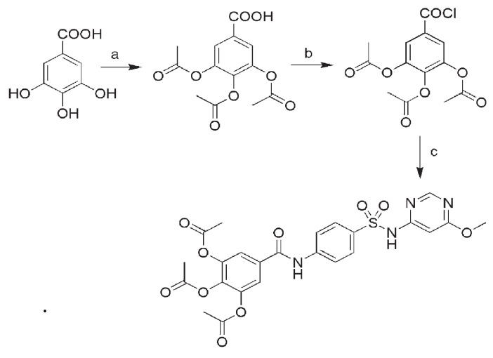
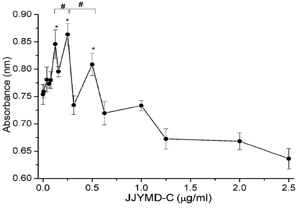
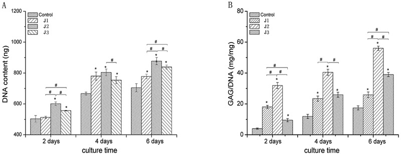
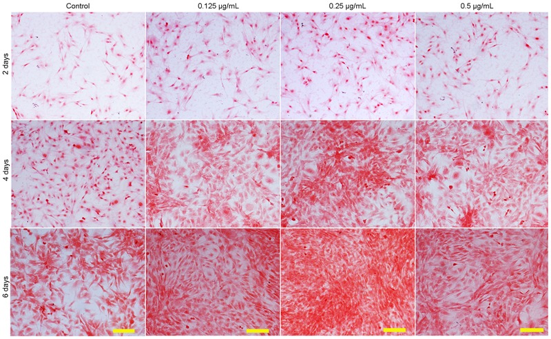
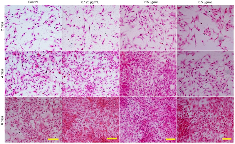
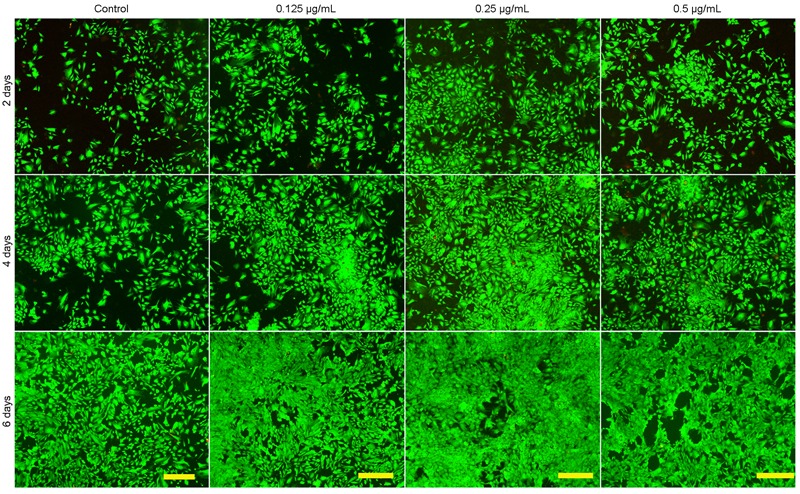
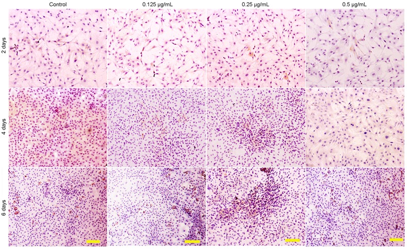
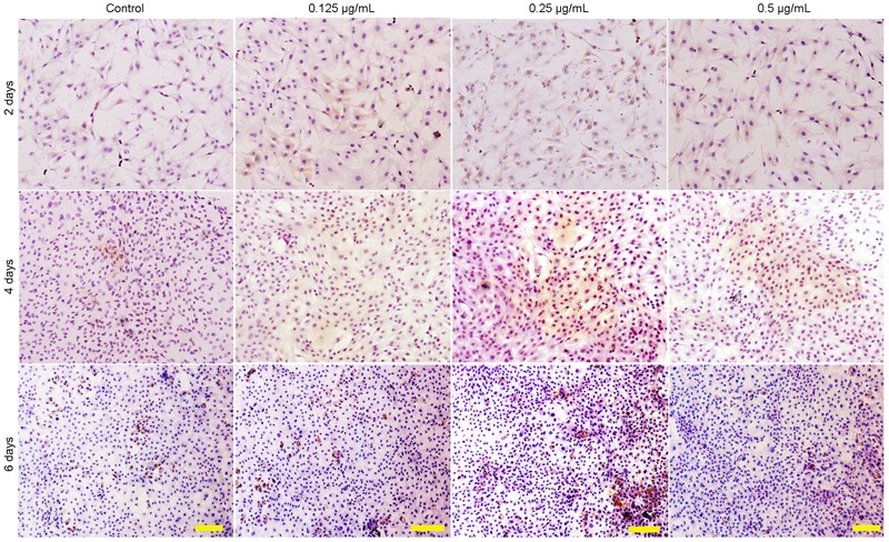
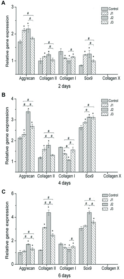
Tissue engineering encapsulated cells such as chondrocytes in the carrier matrix have been widely used to repair cartilage defects. However, chondrocyte phenotype is easily lost when chondrocytes are expanded in vitro by a process defined as "dedifferentiation". To ensure successful therapy, an effective pro-chondrogenic agent is necessary to overcome the obstacle of limited cell numbers in the restoration process, and dedifferentiation is a prerequisite. Gallic acid (GA) has been used in the treatment of arthritis, but its biocompatibility is inferior to that of other compounds. In this study, we modified GA by incorporating sulfamonomethoxine sodium and synthesized a sulfonamido-based gallate, JJYMD-C, and evaluated its effect on chondrocyte metabolism. Our results showed that JJYMD-C could effectively increase the levels of the collagen II, Sox9, and aggrecan genes, promote chondrocyte growth, and enhance secretion and synthesis of cartilage extracellular matrix. On the other hand, expression of the collagen I gene was effectively down-regulated, demonstrating inhibition of chondrocyte dedifferentiation by JJYMD-C. Hypertrophy, as a characteristic of chondrocyte ossification, was undetectable in the JJYMD-C groups. We used JJYMD-C at doses of 0.125, 0.25, and 0.5 µg/mL, and the strongest response was observed with 0.25 µg/mL. This study provides a basis for further studies on a novel agent in the treatment of articular cartilage defects.


Figures

Figure 1
Figure 1. Schematic route and procedures for the synthesis of JJYMD-C. Reagents and conditions: (a) acetyl oxide, oil bath, 120°C; (b) SOCl2, oil bath, 80°C; (c) sulfamonomethoxine sodium, tetrahydrofuran, pyridine, ice bath (0°C).
Figure 2
Figure 2. Cytotoxicity analysis of chondrocytes after 3 days of treatment with different concentrations of JJYMD-C. Data are reported as means±SD for n=20. *P<0.05 compared to control; #P<0.05 comparisons as indicated (one-way ANOVA followed by the Dunnett post hoc test).
Figure 3
Figure 3. Quantification of cell proliferation by detection of DNA content and matrix production by glycosaminoglycan (GAG) analysis. A, Proliferation of chondrocytes cultured in vitro with 0 (control), 0.125 (J1), 0.25 (J2), and 0.5 µg/mL (J3) JJYMD-C for 2, 4, and 6 days. B, GAG level (mg) normalized to DNA content (mg). Data are reported as means±SD for 4 independent experiments *P<0.05 compared to control; #P<0.05 comparisons as indicated (one-way ANOVA followed by the Dunnett post hoc test).
Figure 4
Figure 4. Safranin-O-staining showing the synthesis of extracellular matrix by chondrocytes cultured in vitro with 0 (control), 0.125, 0.25, and 0.5 µg/mL JJYMD-C for 2, 4, and 6 days. Scale bar: 100 µm.
Figure 5
Figure 5. Hematoxylin-eosin staining showing the morphology of chondrocytes cultured in vitro with 0 (control), 0.125, 0.25, and 0.5 µg/mL JJYMD-C for 2, 4, and 6 days. Scale bar: 100 µm.
Figure 6
Figure 6. Laser-scanning confocal microscopy images showing the viability of chondrocytes cultured in vitro with 0 (control), 0.125, 0.25, and 0.5 µg/mL JJYMD-C for 2, 4, and 6 days. Scale bar: 100 µm.
Figure 7
Figure 7. Immunohistochemical staining revealed the presence of type I collagen. Chondrocytes cultured in vitro with 0 (control), 0.125, 0.25, and 0.5 µg/mL JJYMD-C for 2, 4, and 6 days. Scale bar: 100 µm.
Figure 8
Figure 8. Immunohistochemical staining revealed the presence of type II collagen. Chondrocytes cultured in vitro with 0 (control), 0.125, 0.25, and 0.5 µg/mL JJYMD-C for 2, 4, and 6 days. Scale bar: 100 µm.
Figure 9
Figure 9. Quantitative comparison of extracellular matrix-related gene expression by quantitative real-time polymerase chain reaction. The chondrocytes were cultured with 0 (control), 0.125 (J1), 0.25 (J2), and 0.5 µg/mL (J3) JJYMD-C for 2 days (A), 4 days (B), and 6 days (C) (n=3 for each experiment). The levels of gene expression in media containing JJYMD-C were analyzed by the 2-ΔΔCT method, and glyceraldehyde-3-phosphate dehydrogenase was used as internal control. Data are reported as means±SD of three independent culture experiments. *P<0.05 compared to control; #P<0.05 comparisons as indicated (one-way ANOVA followed by the Dunnett post hoc test).

Similar articles

Cited by

References

    1. Menetrey J, Unno-Veith F, Madry H, Van Breuseghem I. Epidemiology and imaging of the subchondral bone in articular cartilage repair. Knee Surg Sports Traumatol Arthrosc. 2010;18:463–471. doi: 10.1007/s00167-010-1053-0. - DOI - PubMed
    1. Carranza-Bencano A, Garcia-Paino L, Armas Padron JR, Cayuela Dominguez A. Neochondrogenesis in repair of full-thickness articular cartilage defects using free autogenous periosteal grafts in the rabbit. A follow-up in six months. Osteoarthritis Cartilage. 2000;8:351–358. doi: 10.1053/joca.1999.0309. - DOI - PubMed
    1. Wakitani S, Imoto K, Yamamoto T, Saito M, Murata N, Yoneda M. Human autologous culture expanded bone marrow mesenchymal cell transplantation for repair of cartilage defects in osteoarthritic knees. Osteoarthritis Cartilage. 2002;10:199–206. doi: 10.1053/joca.2001.0504. - DOI - PubMed
    1. Buckwalter JA, Mankin HJ. Articular cartilage: tissue design and chondrocyte-matrix interactions. Instr Course Lect. 1998;47:477–486. - PubMed
    1. Zhang S, Chen L, Jiang Y, Cai Y, Xu G, Tong T, et al. Bi-layer collagen/microporous electrospun nanofiber scaffold improves the osteochondral regeneration. Acta Biomater. 2013;9:7236–7247. doi: 10.1016/j.actbio.2013.04.003. - DOI - PubMed

Publication types

MeSH terms