

Identification of the augmin complex in the filamentous fungus Aspergillus nidulans
- PMID: 25003582
- PMCID: PMC4086812
- DOI: 10.1371/journal.pone.0101471
Identification of the augmin complex in the filamentous fungus Aspergillus nidulans
Abstract
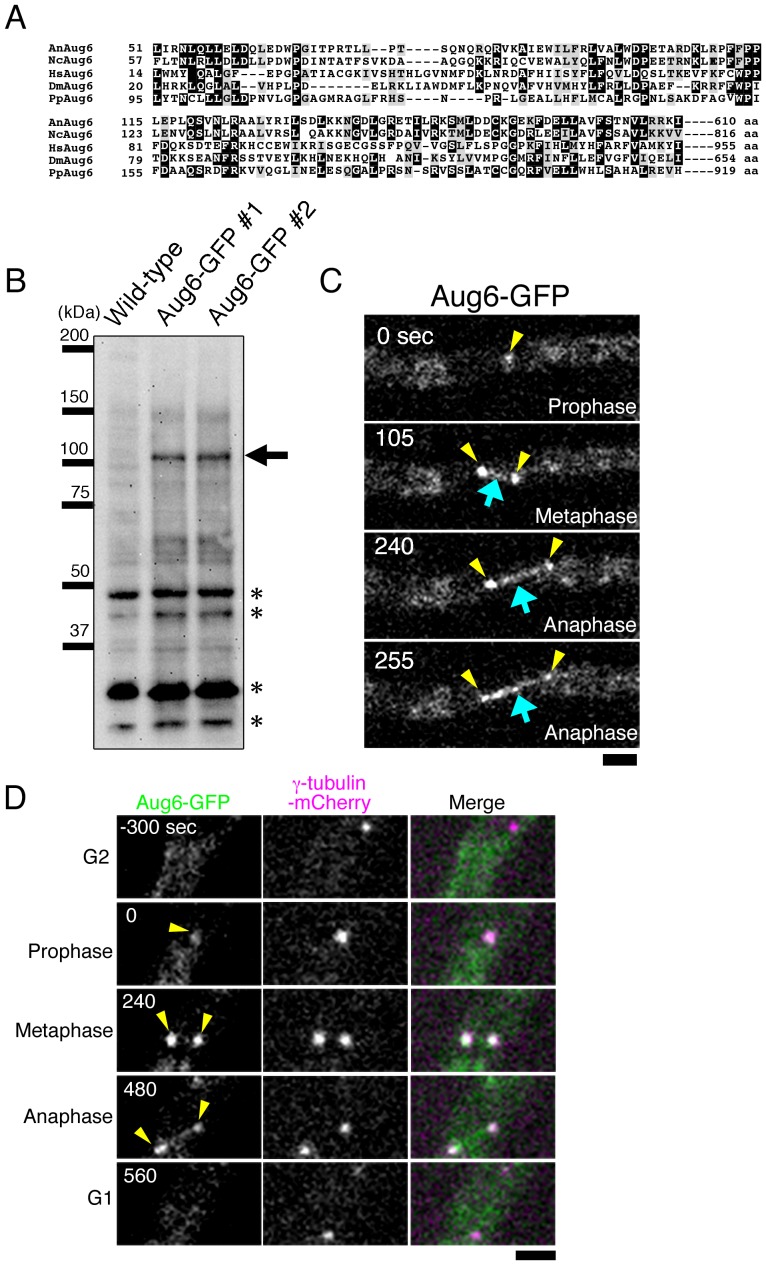
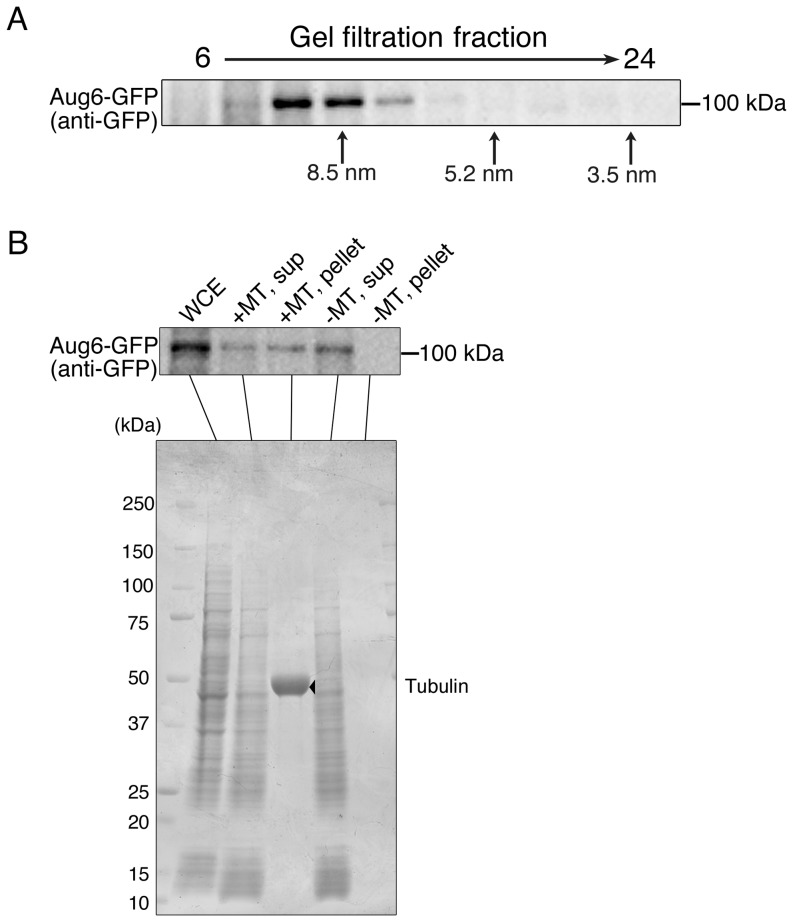
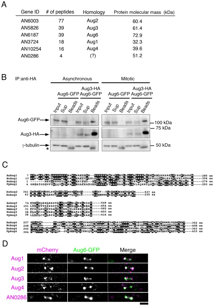
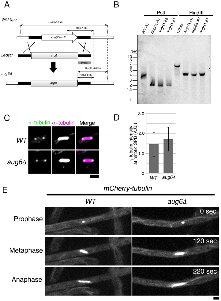
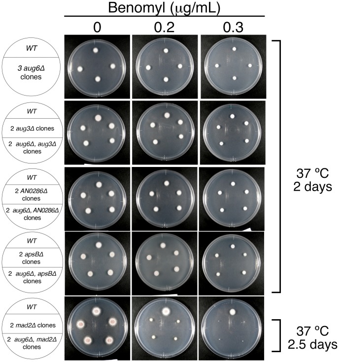
Augmin is a protein complex that binds to spindle microtubules (MTs), recruits the potent MT nucleator, γ-tubulin, and thereby promotes the centrosome-independent MT generation within mitotic and meiotic spindles. Augmin is essential for acentrosomal spindle assembly, which is commonly observed during mitosis in plants and meiosis in female animals. In many animal somatic cells that possess centrosomes, the centrosome- and augmin-dependent mechanisms work cooperatively for efficient spindle assembly and cytokinesis. Yeasts have lost the augmin genes during evolution. It is hypothesized that their robust MT nucleation from the spindle pole body (SPB), the centrosome-equivalent structure in fungi, compensates for the lack of augmin. Intriguingly, however, a gene homologous to an augmin subunit (Aug6/AUGF) has been found in the genome of filamentous fungi, which has the SPB as a robust MT nucleation centre. Here, we aimed to clarify if the augmin complex is present in filamentous fungi and to identify its role in mitosis. By analysing the Aug6-like gene in the filamentous fungus Aspergillus nidulans, we found that it forms a large complex with several other proteins that share weak but significant homology to known augmin subunits. In A. nidulans, augmin was enriched at the SPB and also associated with spindle MTs during mitosis. However, the augmin gene disruptants did not exhibit growth defects under normal, checkpoint-deficient, or MT-destabilised conditions. Moreover, we obtained no evidence that A. nidulans augmin plays a role in γ-tubulin recruitment or in mitotic cell division. Our study uncovered the conservation of the augmin complex in the fungal species, and further suggests that augmin has several functions, besides mitotic spindle MT nucleation, that are yet to be identified.
Conflict of interest statement
Figures
Similar articles
-
How Microtubules Build the Spindle Branch by Branch.Annu Rev Cell Dev Biol. 2022 Oct 6;38:1-23. doi: 10.1146/annurev-cellbio-120420-114559. Epub 2022 Jun 27. Annu Rev Cell Dev Biol. 2022. PMID: 35759800 Free PMC article. Review.
-
The microtubule-associated protein EML3 regulates mitotic spindle assembly by recruiting the Augmin complex to spindle microtubules.J Biol Chem. 2019 Apr 5;294(14):5643-5656. doi: 10.1074/jbc.RA118.007164. Epub 2019 Feb 5. J Biol Chem. 2019. PMID: 30723163 Free PMC article.
-
Characterization of the Arabidopsis augmin complex uncovers its critical function in the assembly of the acentrosomal spindle and phragmoplast microtubule arrays.Plant Cell. 2012 Apr;24(4):1494-509. doi: 10.1105/tpc.112.096610. Epub 2012 Apr 13. Plant Cell. 2012. PMID: 22505726 Free PMC article.
-
Regulation of microtubule-based microtubule nucleation by mammalian polo-like kinase 1.Proc Natl Acad Sci U S A. 2011 Jul 12;108(28):11446-51. doi: 10.1073/pnas.1106223108. Epub 2011 Jun 20. Proc Natl Acad Sci U S A. 2011. PMID: 21690413 Free PMC article.
-
Microtubule nucleation for the assembly of acentrosomal microtubule arrays in plant cells.New Phytol. 2019 Jun;222(4):1705-1718. doi: 10.1111/nph.15705. Epub 2019 Feb 25. New Phytol. 2019. PMID: 30681146 Review.
Cited by
-
How Microtubules Build the Spindle Branch by Branch.Annu Rev Cell Dev Biol. 2022 Oct 6;38:1-23. doi: 10.1146/annurev-cellbio-120420-114559. Epub 2022 Jun 27. Annu Rev Cell Dev Biol. 2022. PMID: 35759800 Free PMC article. Review.
-
Assembly and regulation of γ-tubulin complexes.Open Biol. 2018 Mar;8(3):170266. doi: 10.1098/rsob.170266. Open Biol. 2018. PMID: 29514869 Free PMC article. Review.
-
Horizontal Gene Transfer From Bacteria and Plants to the Arbuscular Mycorrhizal Fungus Rhizophagus irregularis.Front Plant Sci. 2018 May 25;9:701. doi: 10.3389/fpls.2018.00701. eCollection 2018. Front Plant Sci. 2018. PMID: 29887874 Free PMC article.
-
Regulation of microtubule nucleation mediated by γ-tubulin complexes.Protoplasma. 2017 May;254(3):1187-1199. doi: 10.1007/s00709-016-1070-z. Epub 2017 Jan 10. Protoplasma. 2017. PMID: 28074286 Review.
-
Oocyte Meiotic Spindle Assembly and Function.Curr Top Dev Biol. 2016;116:65-98. doi: 10.1016/bs.ctdb.2015.11.031. Epub 2016 Jan 23. Curr Top Dev Biol. 2016. PMID: 26970614 Free PMC article. Review.
References
-
- Moritz M, Agard DA (2001) Gamma-tubulin complexes and microtubule nucleation. Curr Opin Struct Biol 11: 174–181. - PubMed
Publication types
MeSH terms
Substances
LinkOut - more resources
Full Text Sources
Other Literature Sources
