

A microfluidic method to synthesize transferrin-lipid nanoparticles loaded with siRNA LOR-1284 for therapy of acute myeloid leukemia
- PMID: 25003978
- PMCID: PMC4312591
- DOI: 10.1039/c4nr01510j
A microfluidic method to synthesize transferrin-lipid nanoparticles loaded with siRNA LOR-1284 for therapy of acute myeloid leukemia
Abstract
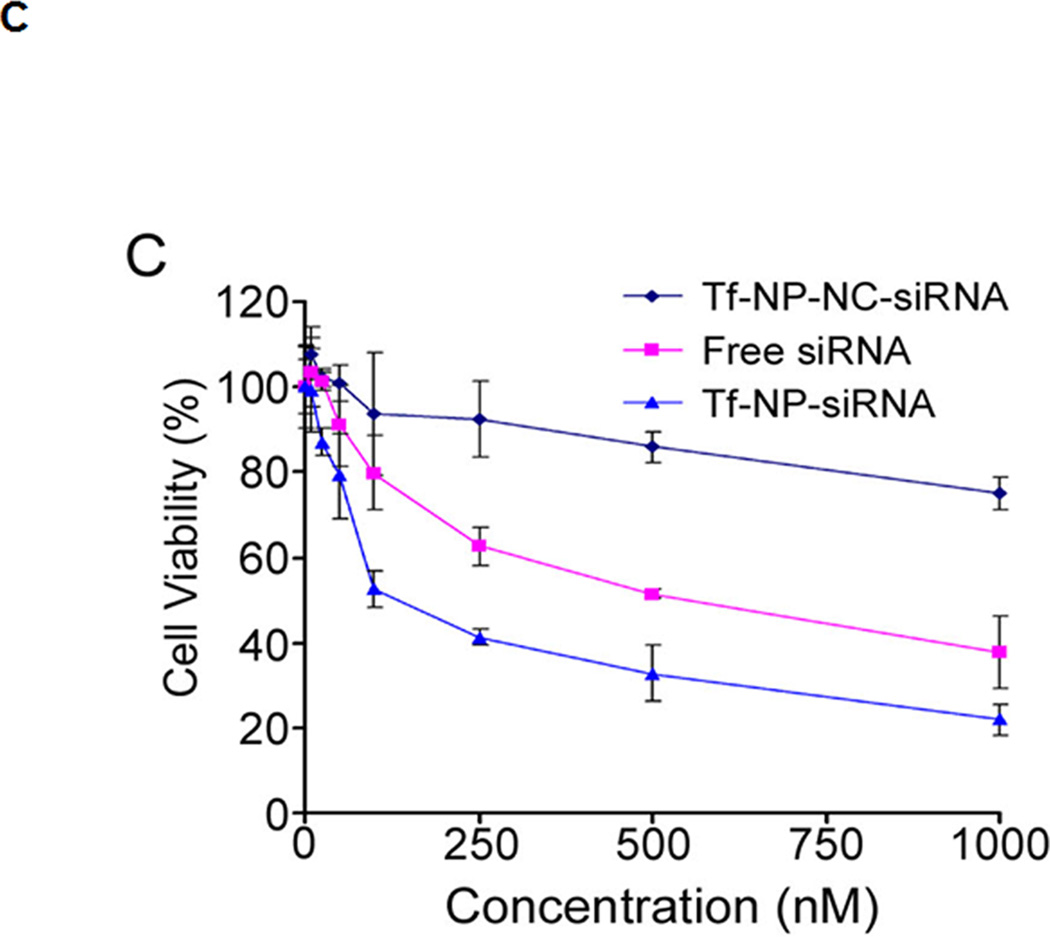
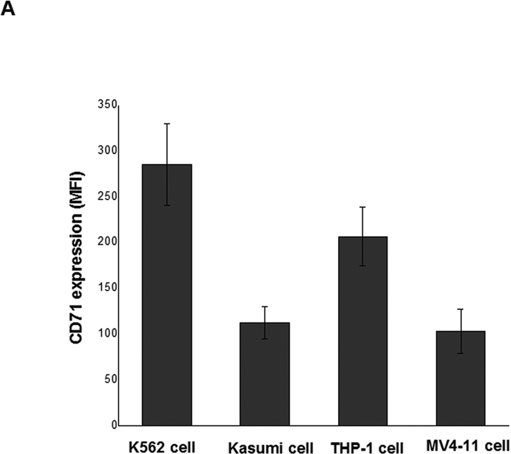
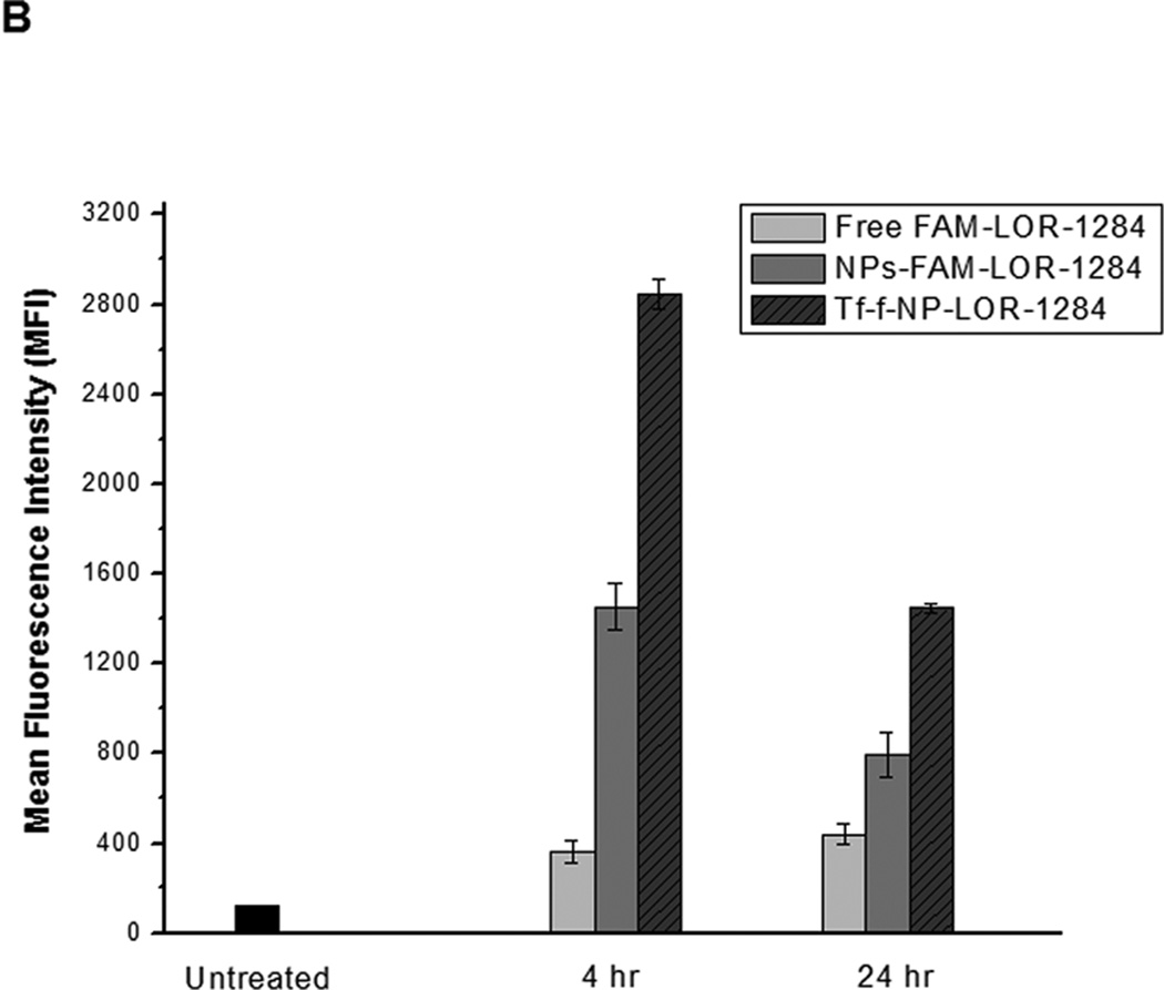
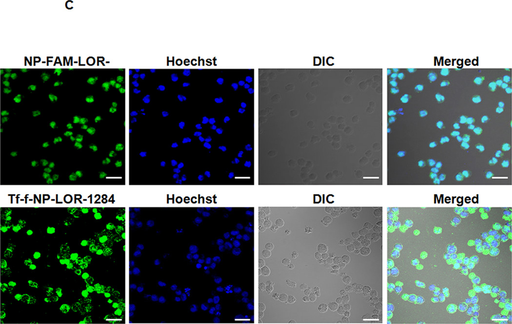
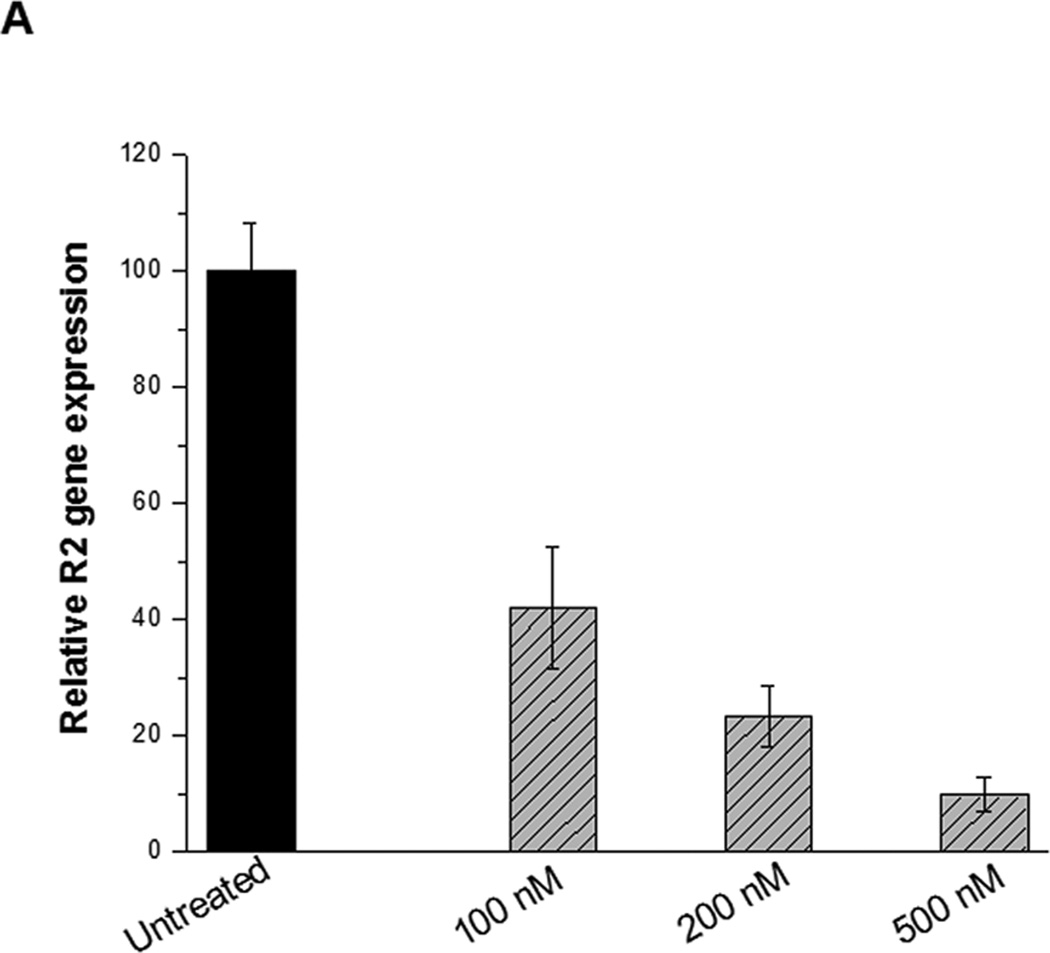
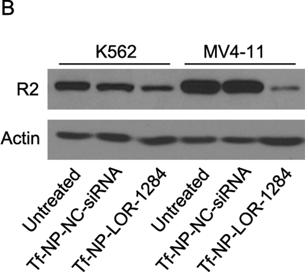
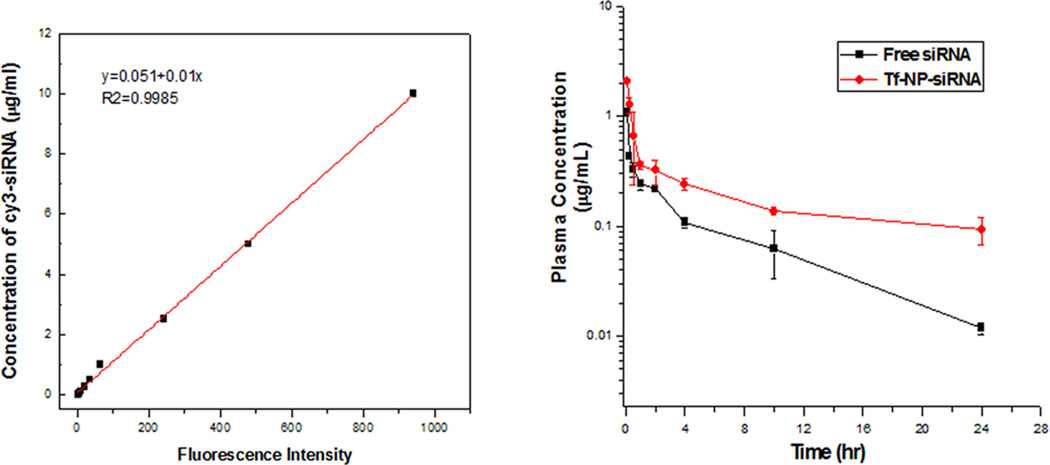
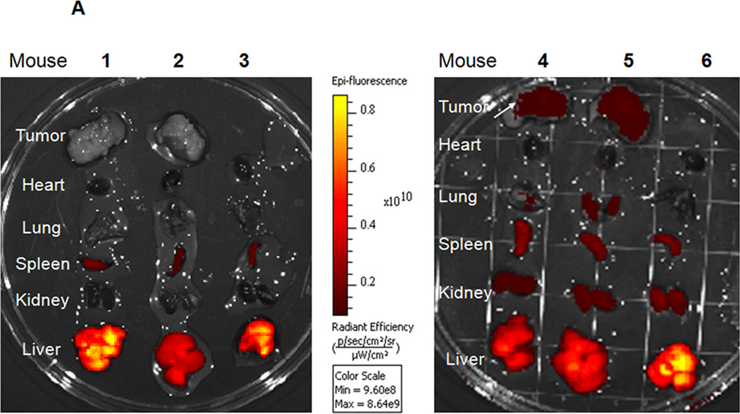
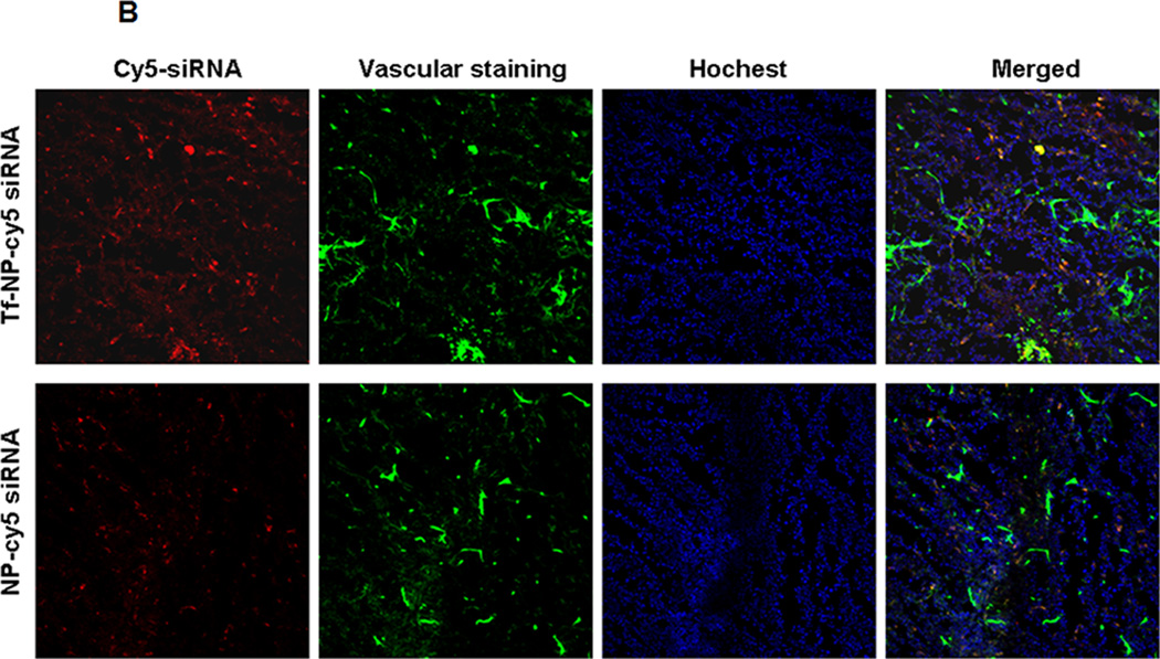
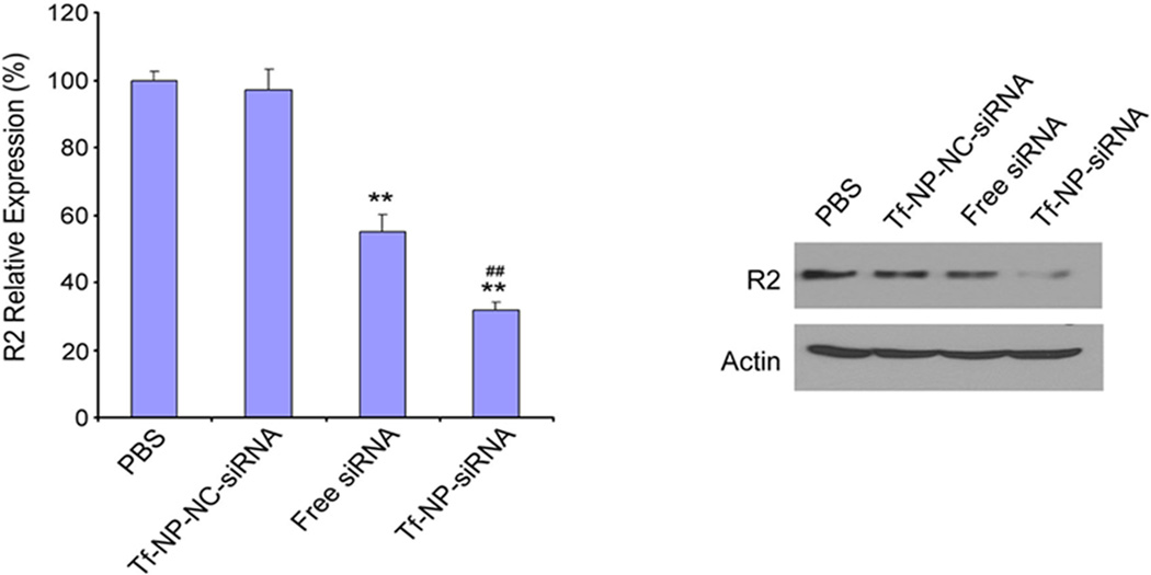
The siRNA LOR-1284 targets the R2 subunit of ribonucleotide reductase (RRM2) and has shown promise in cancer therapy. In this study, transferrin (Tf) conjugated lipid nanoparticles (Tf-NP-LOR-1284) were synthesized by microfluidic hydrodynamic focusing (MHF) and evaluated for the targeted delivery of LOR-1284 siRNA into acute myeloid leukemia (AML) cells. The in vitro study showed that Tf-NP-LOR-1284 can protect LOR-1284 from serum nuclease degradation. Selective uptake of Tf-NP-LOR-1284 was observed in MV4-11 cells. In addition, qRT-PCR and Western blot results revealed that Tf-NP-LOR-1284 was more effective than the free LOR-1284 in reducing the R2 mRNA and protein levels. The Tf-NP-LOR-1284 showed prolonged circulation time and increased AUC after i.v. administration relative to the free LOR-1284. Furthermore, Tf-NP-LOR-1284 facilitated increased accumulation at the tumor site along with the decreased R2 mRNA and protein expression in a murine xenograft model. These results suggest that Tf-conjugated NPs prepared by MHF provide a suitable platform for efficient and specific therapeutic delivery of LOR-1284 into AML cells.
Figures
Similar articles
-
Targeted delivery of microRNA-29b by transferrin-conjugated anionic lipopolyplex nanoparticles: a novel therapeutic strategy in acute myeloid leukemia.Clin Cancer Res. 2013 May 1;19(9):2355-67. doi: 10.1158/1078-0432.CCR-12-3191. Epub 2013 Mar 14. Clin Cancer Res. 2013. PMID: 23493348 Free PMC article.
-
Tumor-targeting transferrin nanoparticles for systemic polymerized siRNA delivery in tumor-bearing mice.Bioconjug Chem. 2013 Nov 20;24(11):1850-60. doi: 10.1021/bc400226b. Epub 2013 Oct 25. Bioconjug Chem. 2013. PMID: 24107100
-
A Polyethylenimine-Containing and Transferrin-Conjugated Lipid Nanoparticle System for Antisense Oligonucleotide Delivery to AML.Biomed Res Int. 2016;2016:1287128. doi: 10.1155/2016/1287128. Epub 2016 Jan 26. Biomed Res Int. 2016. PMID: 27034925 Free PMC article.
-
Targeted delivery of antisense oligodeoxynucleotide by transferrin conjugated pH-sensitive lipopolyplex nanoparticles: a novel oligonucleotide-based therapeutic strategy in acute myeloid leukemia.Mol Pharm. 2010 Feb 1;7(1):196-206. doi: 10.1021/mp900205r. Mol Pharm. 2010. PMID: 19852511 Free PMC article.
-
Novel transferrin modified and doxorubicin loaded Pluronic 85/lipid-polymeric nanoparticles for the treatment of leukemia: In vitro and in vivo therapeutic effect evaluation.Biomed Pharmacother. 2017 Feb;86:547-554. doi: 10.1016/j.biopha.2016.11.121. Epub 2016 Dec 23. Biomed Pharmacother. 2017. PMID: 28024291
Cited by
-
Fc-modified exenatide-loaded nanoparticles for oral delivery to improve hypoglycemic effects in mice.Sci Rep. 2018 Jan 15;8(1):726. doi: 10.1038/s41598-018-19170-y. Sci Rep. 2018. PMID: 29335533 Free PMC article.
-
Microfluidic Manufacture of Lipid-Based Nanomedicines.Pharmaceutics. 2022 Sep 14;14(9):1940. doi: 10.3390/pharmaceutics14091940. Pharmaceutics. 2022. PMID: 36145688 Free PMC article. Review.
-
Role of Four Different Kinds of Polyethylenimines (PEIs) in Preparation of Polymeric Lipid Nanoparticles and Their Anticancer Activity Study.J Cancer. 2016 Apr 28;7(7):872-82. doi: 10.7150/jca.13855. eCollection 2016. J Cancer. 2016. PMID: 27162547 Free PMC article.
-
MicroRNAs as a Novel Tool in the Diagnosis of Liver Lipid Dysregulation and Fatty Liver Disease.Molecules. 2019 Jan 9;24(2):230. doi: 10.3390/molecules24020230. Molecules. 2019. PMID: 30634538 Free PMC article. Review.
-
Microfluidic-Based Single-Cell Study: Current Status and Future Perspective.Molecules. 2018 Sep 13;23(9):2347. doi: 10.3390/molecules23092347. Molecules. 2018. PMID: 30217082 Free PMC article. Review.
References
-
- Morris KV, Chan SW, Jacobsen SE, Looney DJ. Science. 2004;305:1289–1292. - PubMed
-
- Gosselin MA, Guo W, Lee RJ. Bioconjug Chem. 2001;12:989–994. - PubMed
-
- Avolio TM, Lee Y, Feng N, Xiong K, Jin H, Wang M, Vassilakos A, Wright J, Young A. Anticancer Drugs. 2007;18:377–388. - PubMed
-
- Wu M, Sherwin T, Brown WL, Stockley PG. Nanomedicine. 2005;1:67–76. - PubMed
Publication types
MeSH terms
Substances
Grants and funding
LinkOut - more resources
Full Text Sources
Other Literature Sources
Medical
Miscellaneous