Review
. 2014 Jul 7;19(7):9628-54.
doi: 10.3390/molecules19079628.

Ruthenium complexes as NO donors for vascular relaxation induction

Affiliations
Review

Ruthenium complexes as NO donors for vascular relaxation induction

Renata Galvão de Lima et al. Molecules. .

Abstract

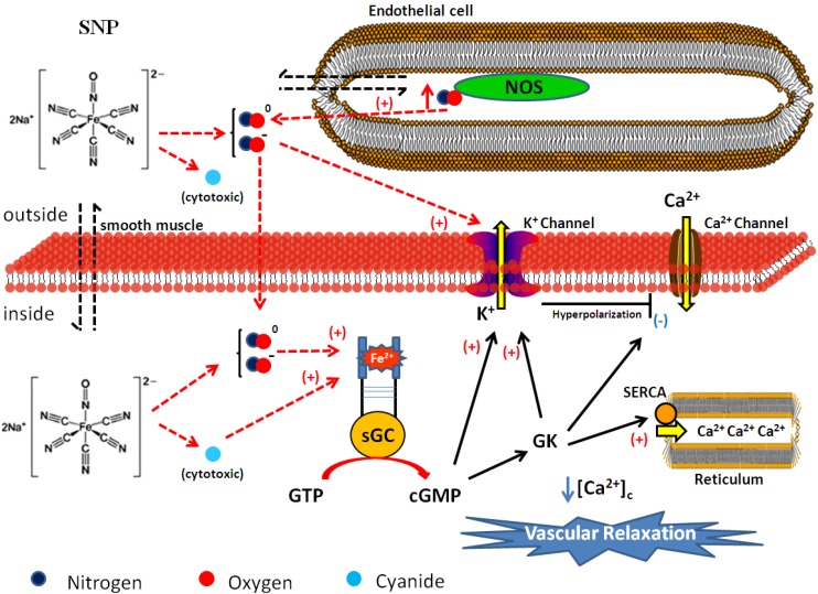
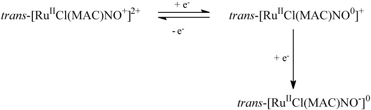
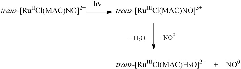
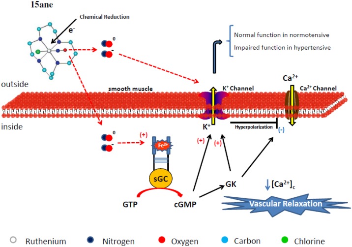
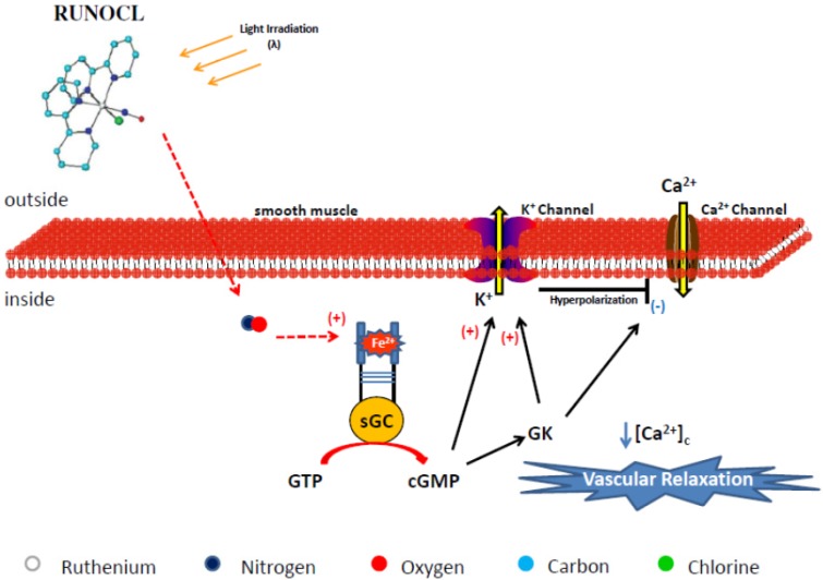
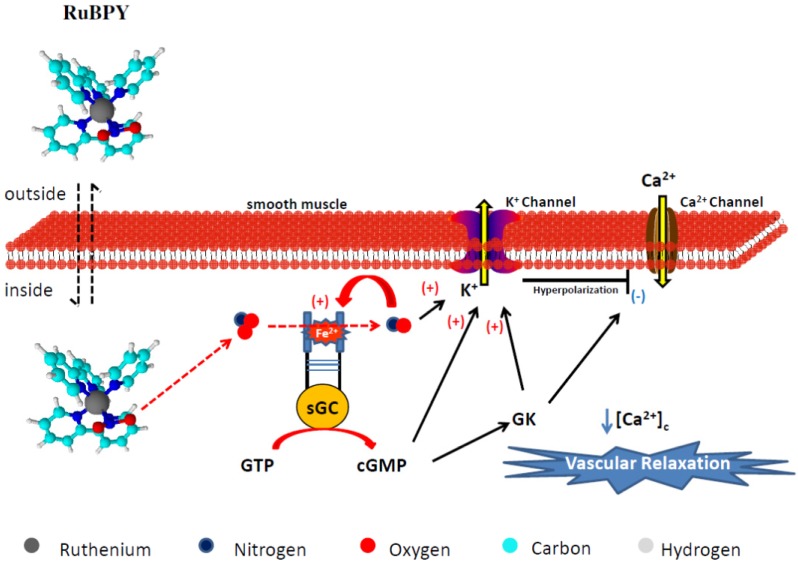
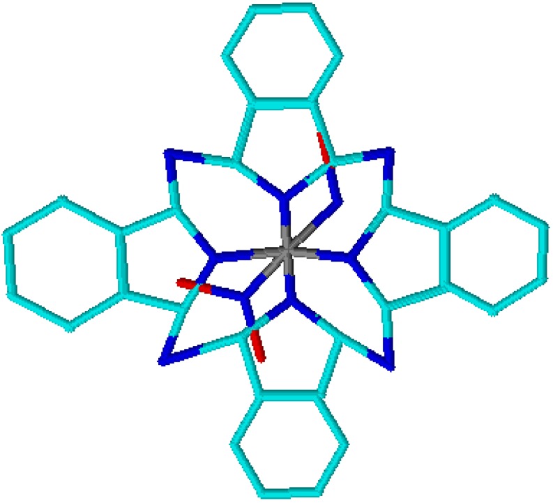
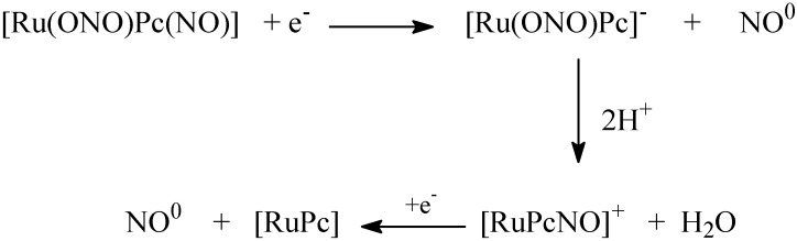
Nitric oxide (NO) donors are substances that can release NO. Vascular relaxation induction is among the several functions of NO, and the administration of NO donors is a pharmacological alternative to treat hypertension. This review will focus on the physicochemical description of ruthenium-derived NO donor complexes that release NO via reduction and light stimulation. In particular, we will discuss the complexes synthesized by our research group over the last ten years, and we will focus on the vasodilation and arterial pressure control elicited by these complexes. Soluble guanylyl cyclase (sGC) and potassium channels are the main targets of the NO species released from the inorganic compounds. We will consider the importance of the chemical structure of the ruthenium complexes and their vascular effects.


Conflict of interest statement

The authors declare that there are no conflicts of interest.

Figures

Figure 1
Figure 1
Proposed nitric oxide release and vasodilating mechanism prompted by sodium nitroprusside (SNP). The colored circles represent atoms in the chemical structure. Legend: NOS = Nitric Oxide Synthase, sGC = soluble guanylyl cyclase, GK = G Kinase Protein, SERCA = sarco/endoplasmic reticulum calcium-ATPase, Ca2+ = calcium, K+ = potassium, [Ca2+]c = cytosolic calcium concentration.
Figure 2
Figure 2
Nitrosyl macrocyclic ruthenium complexes structure. (A) trans-[RuCl([15]ane4)NO]2+ and (B) trans-[RuCl(cyclam)NO]2+. The colored circles represent atoms in the chemical structure (cyan: carbon; blue: nitrogen; green: chlorine; red: oxygen; white: hydrogen and gray: ruthenium).
Scheme 1
Scheme 1
Electrochemical process involved in nitrosyl macrocyclic ruthenium complex.
Scheme 2
Scheme 2
Kinetic process related to the electron reduction of nitrosyl macrocyclic ruthenium complex.
Scheme 3
Scheme 3
General photochemical pathway related to nitrosyl macrocyclic ruthenium complex.
Figure 3
Figure 3
Proposed nitric oxide release and cellular mechanisms involved in the vasodilation promoted by trans-[RuCl(15-aneN4)NO]+ (15ane). The colored circles represent atoms in the chemical structure. The hydrogen atom has been omitted in the structure. Legend: sGC = soluble guanylyl cyclase, GK = G Kinase Protein, Ca2+ = calcium, K+ = potassium, [Ca2+]c = cytosolic calcium concentration.
Figure 4
Figure 4
Chemical structure of polipyridines ruthenium complexes (A) cis-[RuCl(bpy)2NO]2+, (B) cis-[Ru(bpy)2(py)NO]3+ and (C) [Ru(tpy)(NH.NHq)NO]3+. The colored circles represent atoms in the chemical structure (cyan: Carbon; blue: Nitrogen; green: Chlorine; red: Oxygen; white: Hydrogen and gray: Ruthenium).
Figure 5
Figure 5
UV-visible electronic spectra of [RuNO2(tpy)(bpy)]2+ (dashed line) and [Ru(tpy)(bpy)NO]3+ (solid line) in aqueous solution.
Scheme 4
Scheme 4
Interconversion of nitrosyl-nitrite coordinated to ruthenium(II).
Figure 6
Figure 6
Proposed nitric oxide release and vasodilation mechanism elicited by cis-[RuCl(bpy)2(NO)](PF6) (RUNOCL). The colored circles represent atoms in the chemical structure. The hydrogen atom has been omitted in the structure. Legend: sGC = soluble guanylyl cyclase, GK = G Kinase Protein, Ca2+ = calcium, K+ = potassium, [Ca2+]c = cytosolic calcium concentration.
Figure 7
Figure 7
Proposed nitric oxide release and vasodilation mechanism elicited by cis-[RuCl(bpy)2(py)NO2](PF6) (RuBPY). The colored circles represent atoms in the chemical structure. Legend: sGC = soluble guanylyl cyclase, GK = G Kinase Protein, Ca2+ = calcium, K+ = potassium, [Ca2+]c = cytosolic calcium concentration.
Figure 8
Figure 8
Proposed nitric oxide release and vasodilation mechanism induced by Terpy ([Ru(tpy)(NH.NHq)NO]3+). The colored circles represent atoms in the chemical structure. The hydrogen atom has been omitted in the structure. Legend: sGC = soluble guanylyl cyclase, GK = G Kinase Protein, SERCA = sarco/endoplasmic reticulum calcium-ATPase, Ca2+ = calcium, K+ = potassium, [Ca2+]c = cytosolic calcium concentration.
Figure 9
Figure 9
Chemical structure of the nitrosyl phthalocyanine ruthenium complex [Ru(NO)(NO2)pc]. The colored circles represent atoms in the chemical structure (cyan: carbon; blue: nitrogen; red: oxygen and gray: ruthenium).
Scheme 5
Scheme 5
Kinetic process related to the electron reduction of nitrosyl phthalocyanine ruthenium complex.

Similar articles

Cited by

References

    1. Radomski M.W., Palmer R.M.J., Moncada S. The antiaggregating properties of vascular endothelium: Interactions between prostacyclin and nitric oxide. Br. J. Pharmacol. 1987;92:639–646. doi: 10.1111/j.1476-5381.1987.tb11367.x. - DOI - PMC - PubMed
    1. Kline D.D., Yang T.N., Huang P.L., Prabhakar N.R. Altered respiratory responses to hypoxia in mutant mice deficient in neuronal nitric oxide synthase. J. Physiol. 1998;511:273–287. doi: 10.1111/j.1469-7793.1998.273bi.x. - DOI - PMC - PubMed
    1. Bredt D.S., Hwang P.M., Snyder S.H. Localization of nitric oxide synthase indicating a neural role for nitric oxide. Nature. 1990;347:768–770. doi: 10.1038/347768a0. - DOI - PubMed
    1. Garthwaite J. Neural nitric oxide signaling. Trends Neurosci. 1995;18:51–52. doi: 10.1016/0166-2236(95)80014-S. - DOI - PubMed
    1. Akesson B., Lundquist I. Influence of nitric oxide modulators on cholinergically stimulated hormone release from mouse islets. J. Physiol. 1999;515:463–473. doi: 10.1111/j.1469-7793.1999.463ac.x. - DOI - PMC - PubMed

Publication types

MeSH terms