A novel chemopreventive mechanism for a traditional medicine: East Indian sandalwood oil induces autophagy and cell death in proliferating keratinocytes
- PMID: 25004464
- PMCID: PMC4172370
- DOI: 10.1016/j.abb.2014.06.021
A novel chemopreventive mechanism for a traditional medicine: East Indian sandalwood oil induces autophagy and cell death in proliferating keratinocytes
Abstract
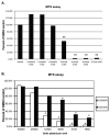
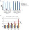
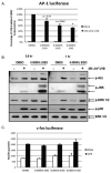
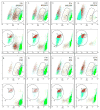
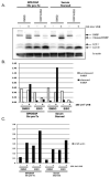
One of the primary components of the East Indian sandalwood oil (EISO) is α-santalol, a molecule that has been investigated for its potential use as a chemopreventive agent in skin cancer. Although there is some evidence that α-santalol could be an effective chemopreventive agent, to date, purified EISO has not been extensively investigated even though it is widely used in cultures around the world for its health benefits as well as for its fragrance and as a cosmetic. In the current study, we show for the first time that EISO-treatment of HaCaT keratinocytes results in a blockade of cell cycle progression as well as a concentration-dependent inhibition of UV-induced AP-1 activity, two major cellular effects known to drive skin carcinogenesis. Unlike many chemopreventive agents, these effects were not mediated through an inhibition of signaling upstream of AP-1, as EISO treatment did not inhibit UV-induced Akt or MAPK activity. Low concentrations of EISO were found to induce HaCaT cell death, although not through apoptosis as annexin V and PARP cleavage were not found to increase with EISO treatment. However, plasma membrane integrity was severely compromised in EISO-treated cells, which may have led to cleavage of LC3 and the induction of autophagy. These effects were more pronounced in cells stimulated to proliferate with bovine pituitary extract and EGF prior to receiving EISO. Together, these effects suggest that EISO may exert beneficial effects upon skin, reducing the likelihood of promotion of pre-cancerous cells to actinic keratosis (AK) and skin cancer.
Keywords: Autophagy; Cell death; Sandalwood oil; Ultraviolet light.
Copyright © 2014 Elsevier Inc. All rights reserved.
Figures

Similar articles
-
Suppression of lipopolysaccharide-stimulated cytokine/chemokine production in skin cells by sandalwood oils and purified α-santalol and β-santalol.Phytother Res. 2014 Jun;28(6):925-32. doi: 10.1002/ptr.5080. Epub 2013 Dec 6. Phytother Res. 2014. PMID: 24318647
-
Skin cancer chemopreventive agent, {alpha}-santalol, induces apoptotic death of human epidermoid carcinoma A431 cells via caspase activation together with dissipation of mitochondrial membrane potential and cytochrome c release.Carcinogenesis. 2005 Feb;26(2):369-80. doi: 10.1093/carcin/bgh325. Epub 2004 Nov 4. Carcinogenesis. 2005. PMID: 15528219
-
East Indian sandalwood (Santalum album L.) oil confers neuroprotection and geroprotection in Caenorhabditis elegans via activating SKN-1/Nrf2 signaling pathway.RSC Adv. 2018 Oct 3;8(59):33753-33774. doi: 10.1039/c8ra05195j. Epub 2018 Oct 2. RSC Adv. 2018. PMID: 30319772 Free PMC article.
-
Medicinal properties of alpha-santalol, a naturally occurring constituent of sandalwood oil: review.Nat Prod Res. 2019 Feb;33(4):527-543. doi: 10.1080/14786419.2017.1399387. Epub 2017 Nov 13. Nat Prod Res. 2019. PMID: 29130352 Review.
-
Anticancer Effects of Sandalwood (Santalum album).Anticancer Res. 2015 Jun;35(6):3137-45. Anticancer Res. 2015. PMID: 26026073 Review.
Cited by
-
Phytosynthesis of silver nanoparticles using aqueous sandalwood (Santalum album L.) leaf extract: Divergent effects of SW-AgNPs on proliferating plant and cancer cells.PLoS One. 2024 Apr 25;19(4):e0300115. doi: 10.1371/journal.pone.0300115. eCollection 2024. PLoS One. 2024. PMID: 38662724 Free PMC article.
-
Autophagy as a targeted therapeutic approach for skin cancer: Evaluating natural and synthetic molecular interventions.Cancer Pathog Ther. 2024 Feb 1;2(4):231-245. doi: 10.1016/j.cpt.2024.01.002. eCollection 2024 Oct. Cancer Pathog Ther. 2024. PMID: 39371094 Free PMC article. Review.
-
Santalol Isomers Inhibit Transthyretin Amyloidogenesis and Associated Pathologies in Caenorhabditis elegans.Front Pharmacol. 2022 Jun 16;13:924862. doi: 10.3389/fphar.2022.924862. eCollection 2022. Front Pharmacol. 2022. PMID: 35784752 Free PMC article.
-
Chemical composition analysis and in vitro biological activities of ten essential oils in human skin cells.Biochim Open. 2017 Apr 26;5:1-7. doi: 10.1016/j.biopen.2017.04.001. eCollection 2017 Dec. Biochim Open. 2017. PMID: 29450150 Free PMC article.
-
Inhibition of Akt Enhances the Chemopreventive Effects of Topical Rapamycin in Mouse Skin.Cancer Prev Res (Phila). 2016 Mar;9(3):215-24. doi: 10.1158/1940-6207.CAPR-15-0419. Epub 2016 Jan 22. Cancer Prev Res (Phila). 2016. PMID: 26801880 Free PMC article.
References
-
- Priest DB. A potent natural antibacterial and anti-inflammatory derived from Australian sandalwood for cosmetic personal use. Cosmetic News. 2004;27:107–109.
-
- Dwivedi C, Abu-Ghazaleh A. Chemopreventive effects of sandalwood oil on skin papillomas in mice. Eur J Cancer Prev. 1997;6:399–401. - PubMed
-
- Dwivedi C, Guan X, Harmsen WL, Voss AL, Goetz-Parten DE, Koopman EM, Johnson KM, Valluri HB, Matthees DP. Chemopreventive effects of alpha-santalol on skin tumor development in CD-1 and SENCAR mice. Cancer epidemiology, biomarkers & prevention: a publication of the American Association for Cancer Research, cosponsored by the American Society of Preventive Oncology. 2003;12:151–156. - PubMed
-
- Dwivedi C, Valluri HB, Guan X, Agarwal R. Chemopreventive effects of alpha-santalol on ultraviolet B radiation-induced skin tumor development in SKH-1 hairless mice. Carcinogenesis. 2006;27:1917–1922. - PubMed
-
- Dwivedi C, Zhang Y. Sandalwood oil prevent skin tumour development in CD1 mice. Eur J Cancer Prev. 1999;8:449–455. - PubMed
Publication types
MeSH terms
Substances
Grants and funding
LinkOut - more resources
Full Text Sources
Other Literature Sources

