Review
. 2014:380:191-212.
doi: 10.1007/978-3-662-43492-5_9.

The molecular basis of IL-10 function: from receptor structure to the onset of signaling

Affiliations
Review

The molecular basis of IL-10 function: from receptor structure to the onset of signaling

Mark R Walter. Curr Top Microbiol Immunol. 2014.

Abstract

Assembly of the cell surface IL-10 receptor complex is the first step in initiating IL-10 signaling pathways that regulate intestinal inflammation, viral persistence and even tumor surveillance. The discovery of IL-10 homologs in the genomes of herpes viruses suggests IL-10 signaling pathways can be manipulated at the level of the receptor complex. This chapter will describe our current molecular understanding of IL-10 receptor assembly based on crystal structures and biochemical analyses of cellular and viral IL-10 receptor complexes.


Figures

Fig. 1
Fig. 1. Sequential Assembly of the IL-10/IL-10R1/IL-10R2 Signaling Complex
a 1:1 IL-10/IL-10R1 complex. b 1:2 IL10/IL-10R1 complex. c 1:2:2 IL-10/IL-10R1/IL-10R2 complex. As noted in the text, an engineered monomeric cIL-10 can induce cell signaling (Josephson et al. 2000). Thus, IL-10R2 can engage the 1:1 IL-10/IL-10R1 complex. The Intracellular receptor domain and JAK/TYK kinases are shown schematically. The triangle represents the docking site/s for unknown proteins required for TNF-α inhibition (Riley et al. 1999). d Enlarged image of one cIL-10 subunit bound to IL-10R1 and IL-10R2 in the ternary signaling complex, which denotes the location of the three binding interfaces that regulate complex formation.
Fig. 2
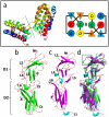
Fig. 2. Structure of cIL-10 and IL-10 Receptors
a Ribbon and schematic diagrams showing the structure and topology of the IL-10 domain swapped dimer. Overall structures of IL-10R1 (b), IL-10R2 (c), and a superposition of the receptor chains (d). Structural differences in the L2 receptor loops are highlighted in the box.
Fig. 3
Fig. 3. Structure of the IL-10/IL-10R1 Binary Complex
a Comparison of the binary cIL-10/IL-10R1 (top) and cmvIL-10/IL-10R1 complexes (bottom). b Enlarged region of the cIL-10/IL-10R1 complex showing the site 1 interface. c Key molecular contacts in the site 1a interface enlarged from Figure 3b. Amino acid sidechains described in the text are shown with carbon atoms and bonds colored yellow, oxygen atoms red, and nitrogen atoms blue. The amino acids are labeled using single letter amino acid codes (e.g. Glu = E). d Important contacts in the site 1b interface, enlarged from Figure 3b. Amino acid side chains described in the text are colored and labeled as described in Figure 3c. e Backbone superposition of site 1 (helices A, B and F) from cIL-10 (green) and cmvIL-10 (blue). cIL-10 and cmvIL-10 residues that donate sidechains to the site 1 interface are colored on the protein backbone yellow and red, respectively. The sidechains of amino acids conserved in the cIL-10 and cmvIL-10 site 1 interface are shown with carbon atoms and bonds colored green for cIL-10 and blue for cmvIL-10. For all sidechains shown, oxygen atoms are red and nitrogen atoms are blue.
Fig. 4
Fig. 4. IL-10R1 Binding Induces Conformational Changes in the IL-10R2 Binding Site (Site 2)
a Orthogonal view, relative to Figure 1b, of the IL-10/IL-10R1 binary complex. b Orthogonal view, relative to Figure 1c, of the IL-10/IL-10R1/IL-10R2 signaling complex derived from a data-driven computational docking strategy (Yoon et al. 2010). c Ribbon diagram of one subunit of IL-10 colored to show the extent of conformational changes observed upon IL-10R1 binding. Distances in the legend correspond to distances between α-carbon atoms in unbound cIL-10 and IL-10R1-bound cIL-10 (Yoon et al. 2006). “Not observed” regions (white) correspond to residues that were not observed in the final electron density maps of unbound cIL-10, but were present in the structure of the cIL-10/sIL-10R1 complex. Residues that form the IL-10R1 binding site 1 are denoted by blue spheres. The sidechains of residues important for IL-10R2 binding are shown in magenta. d Enlargement of the site 2 interface (see Fig. 1d), which shows interactions between aromatic IL-10R2 residues (yellow) and IL-10 residues determined to be important for IL-10R2 binding by mutagenesis (magenta). For all sidechains shown, oxygen atoms are colored red and nitrogen atoms are blue.

Similar articles

Cited by

References

    1. Bandaranayake RM, Ungureanu D, Shan Y, Shaw DE, Silvennoinen O, Hubbard SR. Crystal structures of the JAK2 pseudokinase domain and the pathogenic mutant V617F. Nat Struct Mol Biol. 2012;19:754–9. doi: 10.1038/nsmb.2348. - PMC - PubMed
    1. Bazan JF. Shared architecture of hormone binidng in type I and type II interferon receptors. Cell. 1990;61:753–754. - PubMed
    1. Blackburn SD, Wherry EJ. IL-10, T cell exhaustion and viral persistence. Trends Microbiol. 2007;15:143–6. - PubMed
    1. Bleicher L, de Moura PR, Watanabe L, Colau D, Dumoutier L, Renauld JC, Polikarpov I. Crystal structure of the IL-22/IL-22R1 complex and its implications for the IL-22 signaling mechanism. FEBS Lett. 2008;582:2985–92. doi: 10.1016/j.febslet.2008.07.046. - PubMed
    1. Brooks DG, Trifilo MJ, Edelmann KH, Teyton L, McGavern DB, Oldstone MB. Interleukin-10 determines viral clearance or persistence in vivo. Nat Med. 2006;12:1301–9. - PMC - PubMed

Publication types