. 2014 Jul;70(Pt 7):1790-800.
doi: 10.1107/S1399004714004684. Epub 2014 Jun 24.

Phosphates in the Z-DNA dodecamer are flexible, but their P-SAD signal is sufficient for structure solution

Affiliations

Phosphates in the Z-DNA dodecamer are flexible, but their P-SAD signal is sufficient for structure solution

Zhipu Luo et al. Acta Crystallogr D Biol Crystallogr. 2014 Jul.

Abstract

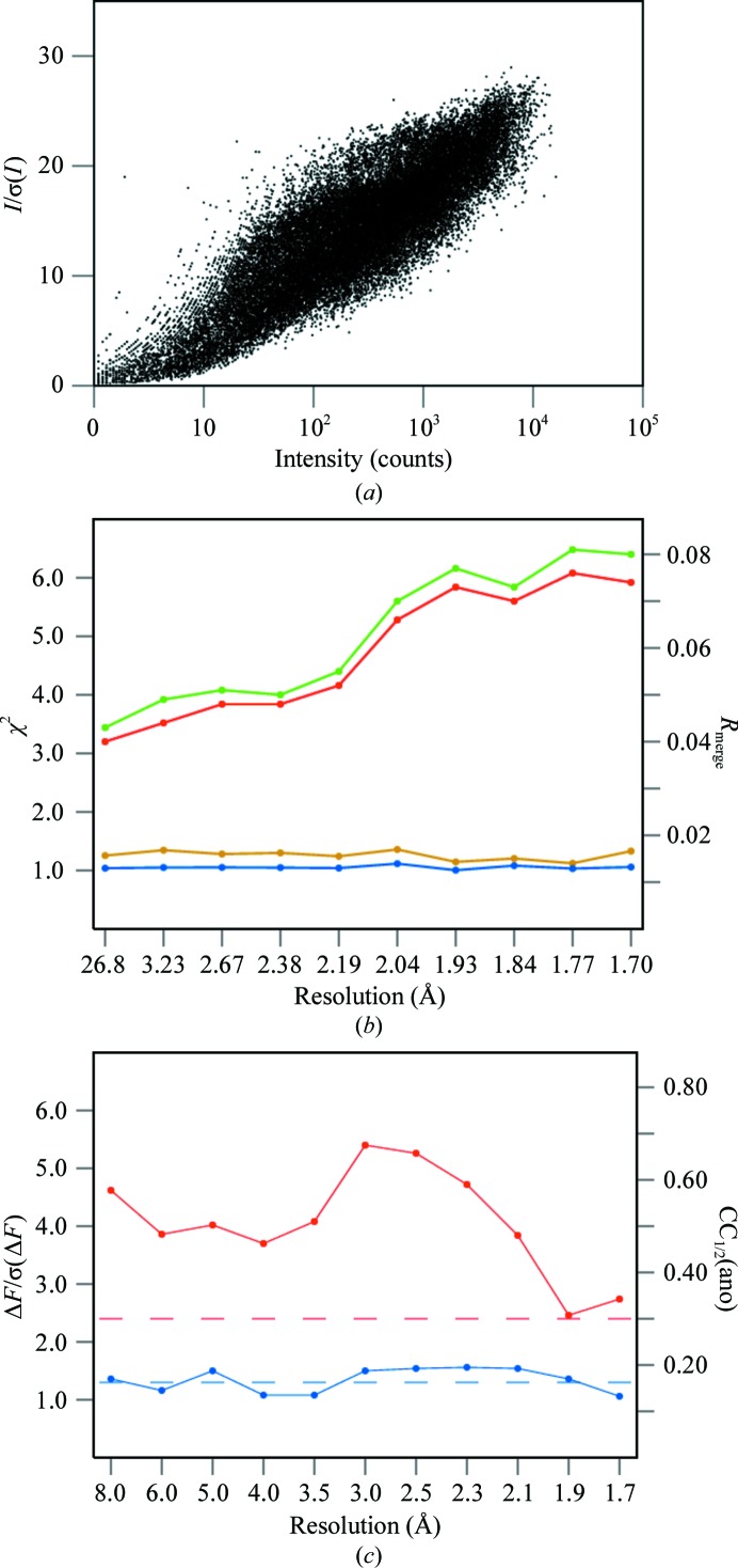
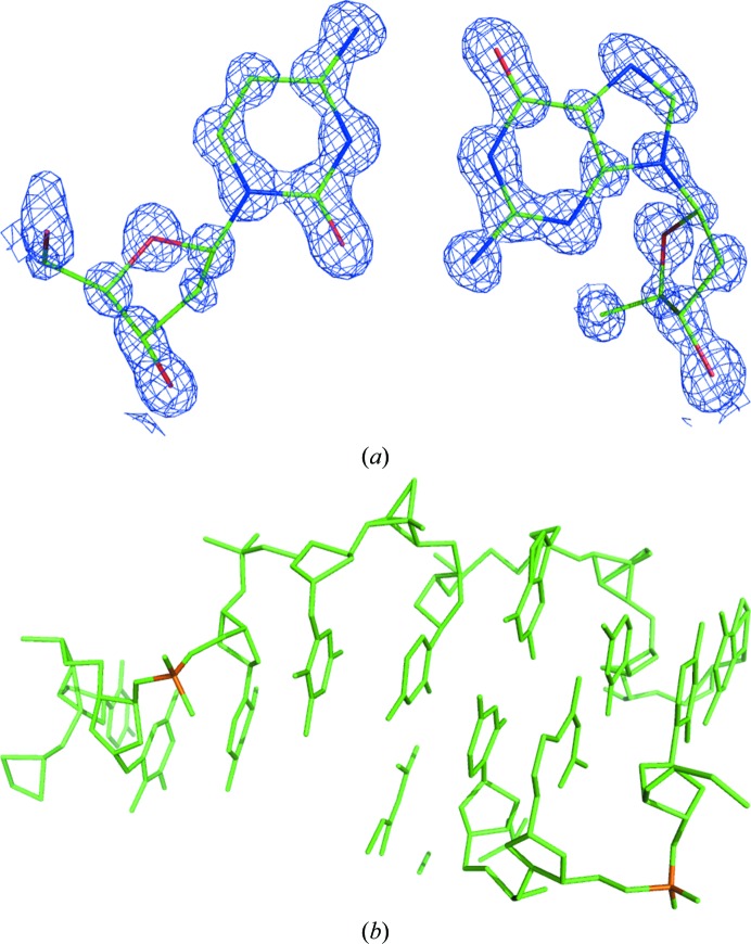
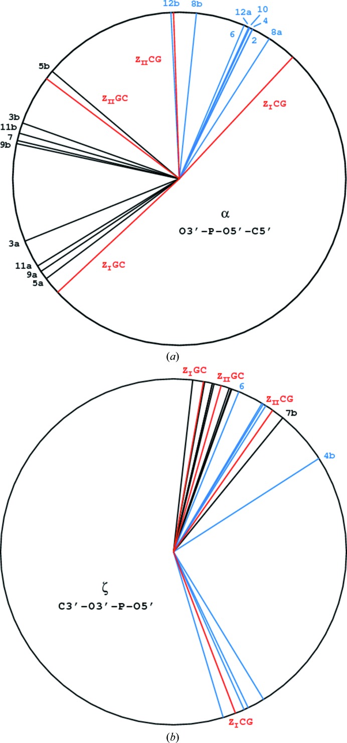
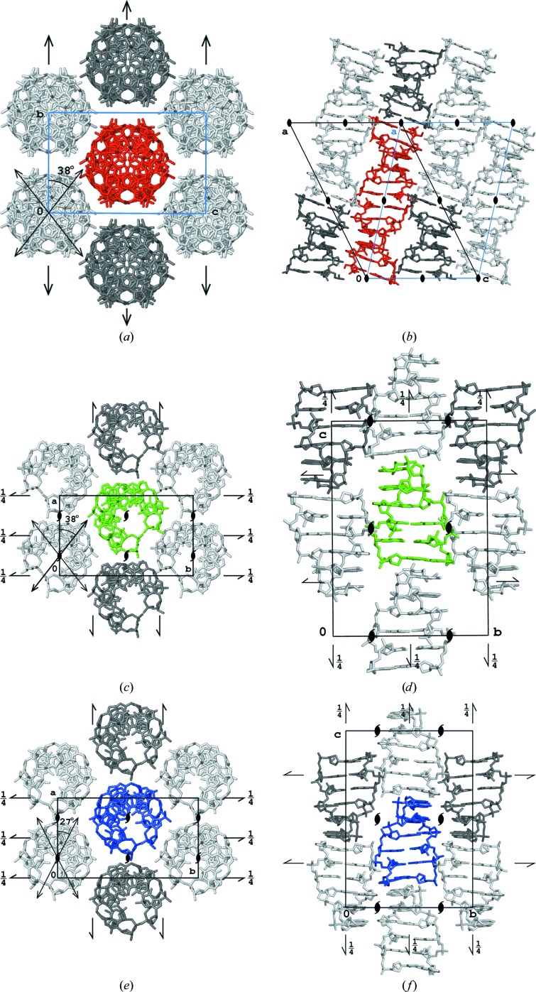
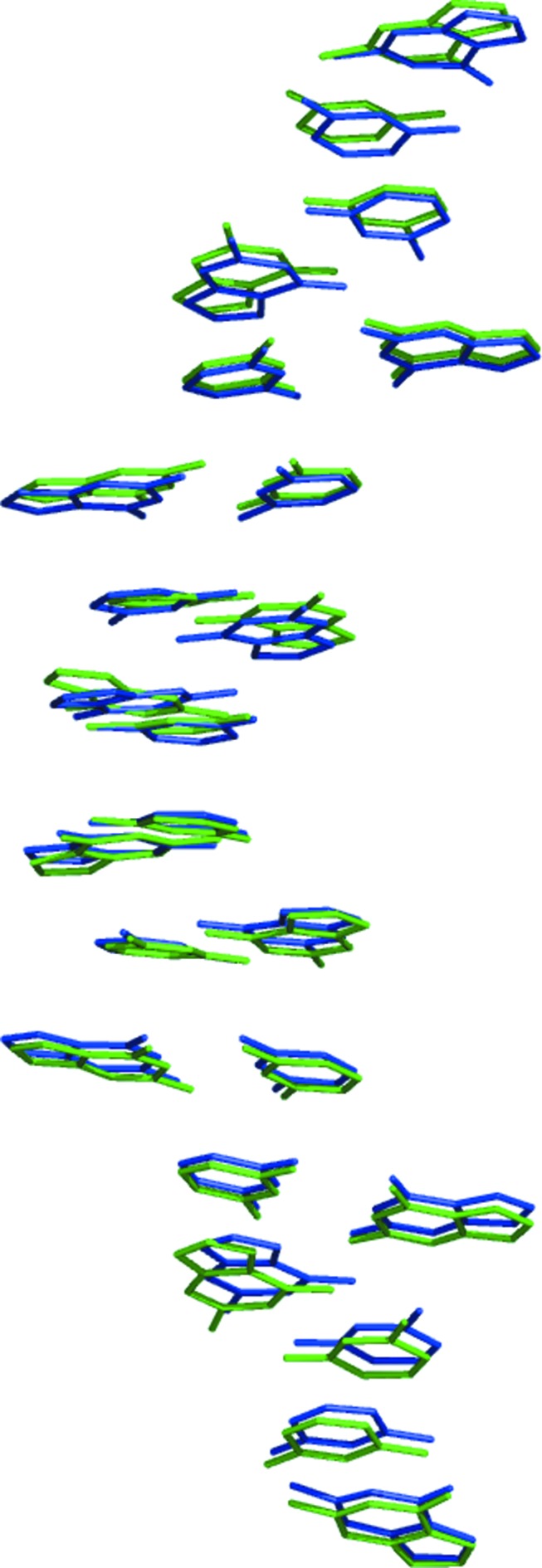
A large number of Z-DNA hexamer duplex structures and a few oligomers of different lengths are available, but here the first crystal structure of the d(CGCGCGCGCGCG)2 dodecameric duplex is presented. Two synchrotron data sets were collected; one was used to solve the structure by the single-wavelength anomalous dispersion (SAD) approach based on the anomalous signal of P atoms, the other set, extending to an ultrahigh resolution of 0.75 Å, served to refine the atomic model to an R factor of 12.2% and an R(free) of 13.4%. The structure consists of parallel duplexes arranged into practically infinitely long helices packed in a hexagonal fashion, analogous to all other known structures of Z-DNA oligomers. However, the dodecamer molecule shows a high level of flexibility, especially of the backbone phosphate groups, with six out of 11 phosphates modeled in double orientations corresponding to the two previously observed Z-DNA conformations: Z(I), with the phosphate groups inclined towards the inside of the helix, and Z(II), with the phosphate groups rotated towards the outside of the helix.

Keywords: Z-DNA dodecamer; Z-DNA structure; flexibility of phosphate groups; phosphorus SAD phasing; ultrahigh resolution.


Figures

Figure 1
Figure 1
The crystal of d(CGCGCGCGCGCG)2 employed to collect both of the diffraction data sets used in this paper.
Figure 2
Figure 2
Various quality criteria of the cg12ano data set. (a) Dependence between signal-to-noise ratio, I/σ(I), and intensity, I, of all reflections. (b) Values of R merge (red and green) and χ2 (blue and yellow) as a function of resolution resulting from treating Friedel mates as equivalent (green and yellow) or independent (red and blue) reflections obtained from SCALEPACK. (c) Anomalous signal to noise, ΔF/σ(ΔF), (blue) and CC1/2(ano) (red) values in resolution bins. The dashed lines show the significance levels of these parameters.
Figure 3
Figure 3
(a) A fragment of the electron-density map at the 1.5σ contour level calculated after P-SAD phasing and density modification by the programs SHELXD and SHELXE, with the final model of a Cyt–Gua pair shown. (b) The model encompassing a total of 243 atoms of the dodecamer obtained from direct-methods phasing by SHELXD (some fragments belong to symmetry-equivalent molecules).
Figure 4
Figure 4
Representations of the individual phosphate groups in either single or double conformations for the CG stage (a) and the GC stage (b) of the dodecamer backbone.
Figure 5
Figure 5
Torsion angles around the backbone bonds involving a P atom (a) for the α bond (O3′—P—O5′—C5′) and (b) for the ζ bond (C3′—O3′—P—O5′). The canonical values for the ZI and ZII phosphate conformations (Saenger, 1983 ▶) are shown in red, the bonds in the CG stage in black and those in the GC stage in blue.
Figure 6
Figure 6
The standard uncertainties estimated from the FMLS refinement for all non-disordered atoms in the dodecamer as a function of their B factors. C atoms are shown in black, N atoms in blue and O atoms in red.
Figure 7
Figure 7
Packing of molecules in the structures of (a, b) the dodecamer, (c, d) hexamer 3p4j and (e, f) hexamer 1i0t viewed along the helices and along the perpendicular direction parallel to the shortest edge of the corresponding unit cell. For the dodecamer, the standard monoclinic C-centered cell is shown in black and the nonstandard monoclinic I-centered cell is shown in blue.
Figure 8
Figure 8
The 12 base pairs of the dodecamer (green) and from two of the 3p4j hexamer duplexes (blue) superimposed onto each other.

Similar articles

Cited by

References

    1. Atwell, S., Meggers, E., Spraggon, G. & Schultz, P. G. (2001). J. Am. Chem. Soc. 123, 12364–12367. - PubMed
    1. Ban, C., Ramakrishnan, B. & Sundaralingam, M. (1996). Biophys. J. 71, 1215–1221. - PMC - PubMed
    1. Bancroft, D., Williams, L. D., Rich, A. & Egli, M. (1994). Biochemistry, 33, 1073–1086. - PubMed
    1. Berman, H. M., Olson, W. K., Beveridge, D. L., Westbrook, J., Gelbin, A., Demeny, T., Hsieh, S. H., Srinivasan, A. R. & Schneider, B. (1992). Biophys. J. 63, 751–759. - PMC - PubMed
    1. Berman, H. M., Westbrook, J., Feng, Z., Gilliland, G., Bhat, T. N., Weissig, H., Shindyalov, I. N. & Bourne, P. E. (2000). Nucleic Acids Res. 28, 235–242. - PMC - PubMed

Publication types

Associated data