. 2014 Jul;70(Pt 7):1801-11.
doi: 10.1107/S139900471400830X. Epub 2014 Jun 24.

Bacterial periplasmic sialic acid-binding proteins exhibit a conserved binding site

Affiliations

Bacterial periplasmic sialic acid-binding proteins exhibit a conserved binding site

Thanuja Gangi Setty et al. Acta Crystallogr D Biol Crystallogr. 2014 Jul.

Abstract

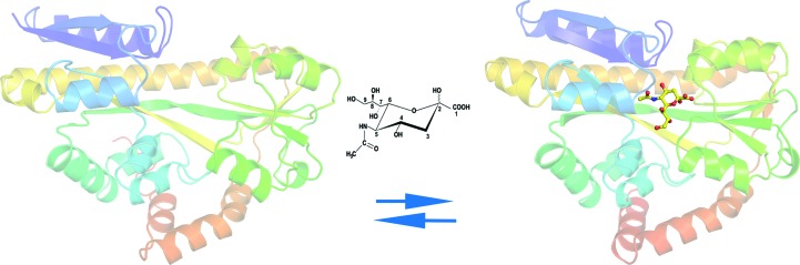
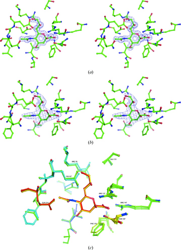
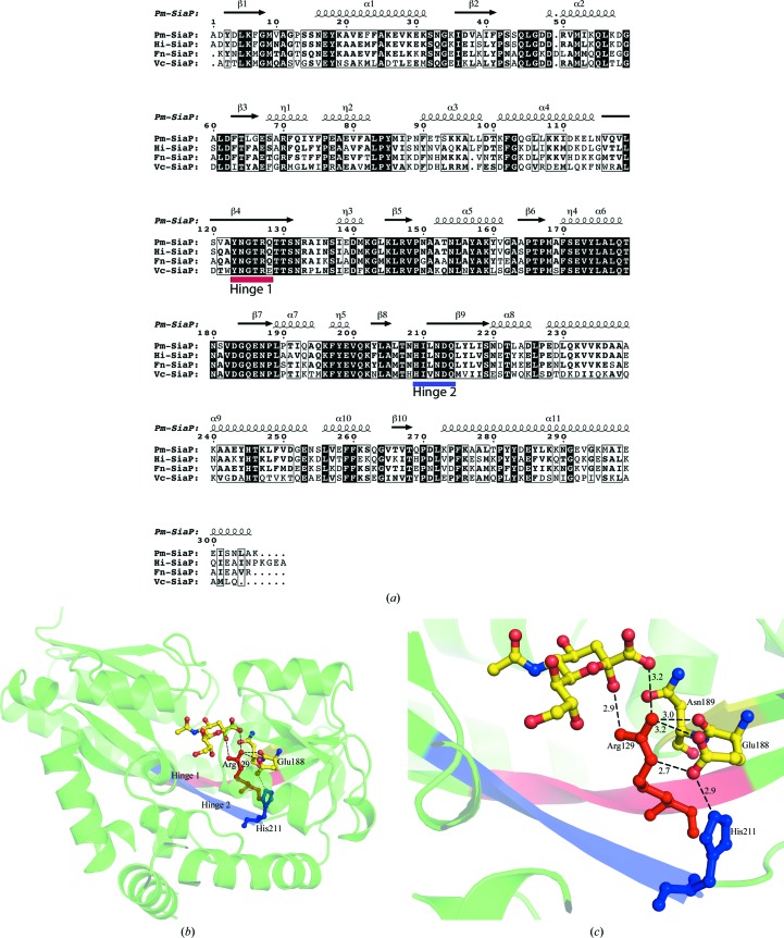
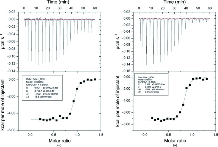
Sialic acids are a family of related nine-carbon sugar acids that play important roles in both eukaryotes and prokaryotes. These sialic acids are incorporated/decorated onto lipooligosaccharides as terminal sugars in multiple bacteria to evade the host immune system. Many pathogenic bacteria scavenge sialic acids from their host and use them for molecular mimicry. The first step of this process is the transport of sialic acid to the cytoplasm, which often takes place using a tripartite ATP-independent transport system consisting of a periplasmic binding protein and a membrane transporter. In this paper, the structural characterization of periplasmic binding proteins from the pathogenic bacteria Fusobacterium nucleatum, Pasteurella multocida and Vibrio cholerae and their thermodynamic characterization are reported. The binding affinities of several mutations in the Neu5Ac binding site of the Haemophilus influenzae protein are also reported. The structure and the thermodynamics of the binding of sugars suggest that all of these proteins have a very well conserved binding pocket and similar binding affinities. A significant conformational change occurs when these proteins bind the sugar. While the C1 carboxylate has been identified as the primary binding site, a second conserved hydrogen-bonding network is involved in the initiation and stabilization of the conformational states.

Keywords: molecular mimicry; sialic acid-binding proteins; structure binding.


Figures

Figure 1
Figure 1
A cartoon diagram showing the unliganded form of Vc-SiaP (in the absence of Neu5Ac) and the ligand-bound form of Fn-SiaP in the presence of Neu5Ac. All structural figures were produced using PyMOL (Schrödinger). The structures are coloured as a gradient from blue to red, with the N-­terminus in blue. The figure clearly shows the closure of the domains around the bound ligand (Neu5Ac), which is shown in ball-and-stick representation.
Figure 2
Figure 2
(a) Cross-eyed stereo diagram of the bound Neu5Ac and the residues around it in the structure of Pm-SiaP. The electron-density map is a 2F oF c map at 1.5 times the r.m.s. deviation. The map was calculated at the end of the refinement. (b) Cross-eyed stereo diagram of the bound Neu5Ac and the residues around it in the structure of Fn-SiaP. The electron-density map is a 2F oF c map at 1.5 times the r.m.s. deviation. The map was calculated at the end of the refinement. (c) A stick diagram showing Neu5Ac and the conserved interacting residues of Hi-SiaP (magenta), Pm-SiaP (blue) and Fn-Siap (cyan). The numbering corresponds to Hi-SiaP.
Figure 3
Figure 3
(a) Sequence alignment of sialic acid-binding proteins from four different pathogenic bacteria using ClustalW2 (EMBL–EBI) and ESPript. The hinge regions are marked and labelled. Hinge 1 in shown in red and hinge 2 in blue. (b) Structural view of the conserved hinge regions of Fn-SiaP, looking down from the Neu5Ac. Hinge 1 and hinge 2 are coloured red and blue, respectively. Arg129 and His211 are also marked with the corresponding hinge colours. (c) Close-up view of conserved residues from the hinge region of Pm-SiaP in the presence of Neu5Ac. The dotted lines show residues interacting by hydrogen/ionic bonds and the distances are labelled in Å.
Figure 4
Figure 4
Calorimetric titration isotherms of Pm-SiaP binding to Neu5Ac (a) and Neu5Gc (b). The top half show the heat change after every injection and the bottom part shows the fit of the curve. Molar ratio is the stoichiometry of the observed binding between the protein and the ligand.

Similar articles

Cited by

References

    1. Adams, P. D. et al. (2011). Methods, 55, 94–106. - PubMed
    1. Almagro-Moreno, S. & Boyd, E. F. (2009). Infect. Immun. 77, 3807–3816. - PMC - PubMed
    1. Chowdhury, N., Norris, J., McAlister, E., Lau, S. Y. K., Thomas, G. H. & Boyd, E. F. (2012). Microbiology, 158, 2158–2167. - PubMed
    1. Dam, J. & Schuck, P. (2005). Biophys. J. 89, 651–666. - PMC - PubMed
    1. Emsley, P., Lohkamp, B., Scott, W. G. & Cowtan, K. (2010). Acta Cryst. D66, 486–501. - PMC - PubMed

Publication types