

Structural basis of the heterodimerization of the MST and RASSF SARAH domains in the Hippo signalling pathway
- PMID: 25004971
- PMCID: PMC4089488
- DOI: 10.1107/S139900471400947X
Structural basis of the heterodimerization of the MST and RASSF SARAH domains in the Hippo signalling pathway
Abstract
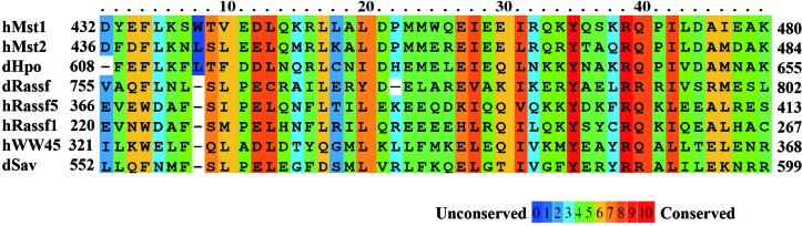
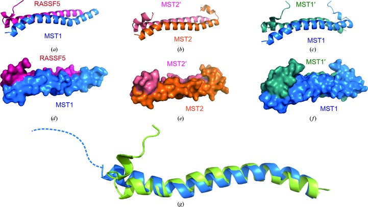
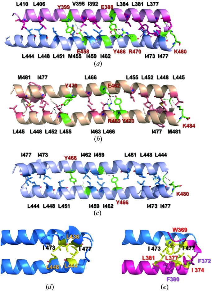
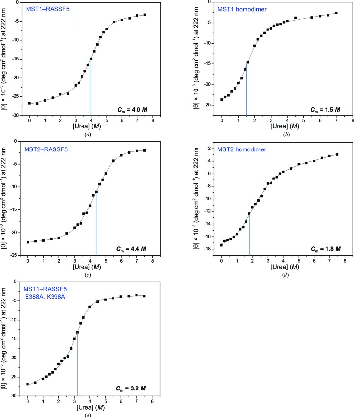
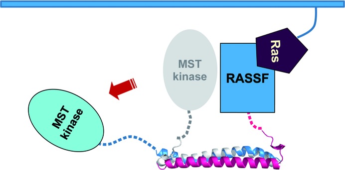
Despite recent progress in research on the Hippo signalling pathway, the structural information available in this area is extremely limited. Intriguingly, the homodimeric and heterodimeric interactions of mammalian sterile 20-like (MST) kinases through the so-called `SARAH' (SAV/RASSF/HPO) domains play a critical role in cellular homeostasis, dictating the fate of the cell regarding cell proliferation or apoptosis. To understand the mechanism of the heterodimerization of SARAH domains, the three-dimensional structures of an MST1-RASSF5 SARAH heterodimer and an MST2 SARAH homodimer were determined by X-ray crystallography and were analysed together with that previously determined for the MST1 SARAH homodimer. While the structure of the MST2 homodimer resembled that of the MST1 homodimer, the MST1-RASSF5 heterodimer showed distinct structural features. Firstly, the six N-terminal residues (Asp432-Lys437), which correspond to the short N-terminal 3₁₀-helix h1 kinked from the h2 helix in the MST1 homodimer, were disordered. Furthermore, the MST1 SARAH domain in the MST1-RASSF5 complex showed a longer helical structure (Ser438-Lys480) than that in the MST1 homodimer (Val441-Lys480). Moreover, extensive polar and nonpolar contacts in the MST1-RASSF5 SARAH domain were identified which strengthen the interactions in the heterodimer in comparison to the interactions in the homodimer. Denaturation experiments performed using urea also indicated that the MST-RASSF heterodimers are substantially more stable than the MST homodimers. These findings provide structural insights into the role of the MST1-RASSF5 SARAH domain in apoptosis signalling.
Keywords: Hippo signalling pathway; MST; RASSF; SARAH domains.
Figures
Similar articles
-
Structural insight into dimeric interaction of the SARAH domains from Mst1 and RASSF family proteins in the apoptosis pathway.Proc Natl Acad Sci U S A. 2007 May 29;104(22):9236-41. doi: 10.1073/pnas.0610716104. Epub 2007 May 21. Proc Natl Acad Sci U S A. 2007. PMID: 17517604 Free PMC article.
-
The dynamic mechanism of RASSF5 and MST kinase activation by Ras.Phys Chem Chem Phys. 2017 Mar 1;19(9):6470-6480. doi: 10.1039/c6cp08596b. Phys Chem Chem Phys. 2017. PMID: 28197608 Free PMC article.
-
Structure of MST2 SARAH domain provides insights into its interaction with RAPL.J Struct Biol. 2014 Mar;185(3):366-74. doi: 10.1016/j.jsb.2014.01.008. Epub 2014 Jan 25. J Struct Biol. 2014. PMID: 24468289
-
RASSF5: An MST activator and tumor suppressor in vivo but opposite in vitro.Curr Opin Struct Biol. 2016 Dec;41:217-224. doi: 10.1016/j.sbi.2016.09.001. Epub 2016 Sep 17. Curr Opin Struct Biol. 2016. PMID: 27643882 Review.
-
Regulation of MST complexes and activity via SARAH domain modifications.Biochem Soc Trans. 2021 Apr 30;49(2):675-683. doi: 10.1042/BST20200559. Biochem Soc Trans. 2021. PMID: 33860801 Free PMC article. Review.
Cited by
-
New type of interaction between the SARAH domain of the tumour suppressor RASSF1A and its mitotic kinase Aurora A.Sci Rep. 2019 Apr 3;9(1):5550. doi: 10.1038/s41598-019-41972-x. Sci Rep. 2019. PMID: 30944388 Free PMC article.
-
Salvador has an extended SARAH domain that mediates binding to Hippo kinase.J Biol Chem. 2018 Apr 13;293(15):5532-5543. doi: 10.1074/jbc.RA117.000923. Epub 2018 Mar 8. J Biol Chem. 2018. PMID: 29519817 Free PMC article.
-
Immunization with Plant-Derived Multimeric H5 Hemagglutinins Protect Chicken against Highly Pathogenic Avian Influenza Virus H5N1.Vaccines (Basel). 2020 Oct 9;8(4):593. doi: 10.3390/vaccines8040593. Vaccines (Basel). 2020. PMID: 33050224 Free PMC article.
-
The components and regulation of the Hippo pathway and its relationships with the progression and treatment of Non-small cell lung cancer (NSCLC).Cancer Cell Int. 2025 Aug 20;25(1):309. doi: 10.1186/s12935-025-03946-0. Cancer Cell Int. 2025. PMID: 40830878 Free PMC article. Review.
-
Regulation of mammalian Ste20 (Mst) kinases.Trends Biochem Sci. 2015 Mar;40(3):149-56. doi: 10.1016/j.tibs.2015.01.001. Epub 2015 Feb 6. Trends Biochem Sci. 2015. PMID: 25665457 Free PMC article. Review.
References
Publication types
MeSH terms
Substances
Associated data
- Actions
- Actions
LinkOut - more resources
Full Text Sources
Other Literature Sources
Research Materials
Miscellaneous
