Review
. 2014 Jul 8:15:26.
doi: 10.1186/1471-2121-15-26.

Recent advances in live cell imaging of hepatoma cells

Affiliations
Review

Recent advances in live cell imaging of hepatoma cells

Sandeep Salipalli et al. BMC Cell Biol. .

Abstract

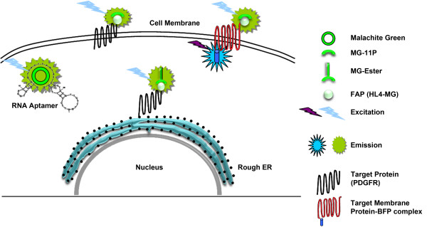
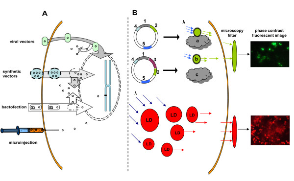
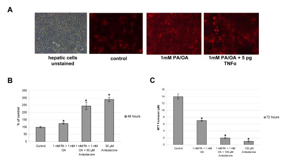
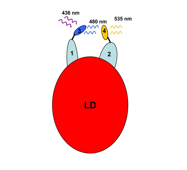
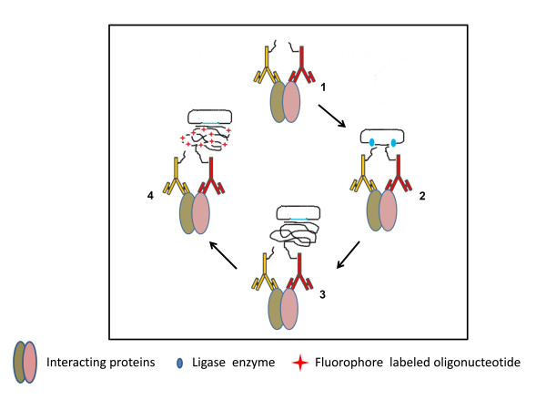
Live cell imaging enables the study of dynamic processes of living cells in real time by use of suitable reporter proteins and the staining of specific cellular structures and/or organelles. With the availability of advanced optical devices and improved cell culture protocols it has become a rapidly growing research methodology. The success of this technique relies mainly on the selection of suitable reporter proteins, construction of recombinant plasmids possessing cell type specific promoters as well as reliable methods of gene transfer. This review aims to provide an overview of the recent developments in the field of marker proteins (bioluminescence and fluorescent) and methodologies (fluorescent resonance energy transfer, fluorescent recovery after photobleaching and proximity ligation assay) employed as to achieve an improved imaging of biological processes in hepatoma cells. Moreover, different expression systems of marker proteins and the modes of gene transfer are discussed with emphasis on the study of lipid droplet formation in hepatocytes as an example.


Figures

Figure 1
Figure 1
Live cell imaging with fluorogen activating proteins (FAP). Malachite green (MG), an organic dye and its membrane permeable (MG-Ester) as well as the membrane impermeable variants (MG-11P) are used in FAP imaging. Essentially, the FAP system consists of an encoded protein such as a single chain antibody fragments to activate the fluorescent dye and/or its derivatives. Note, initially MG was used to visualize RNA aptamers. The intracellular trafficking of the membrane bound receptor tyrosine kinase PDGFR is given as an example. Similarly, membrane bound proteins tagged with both FAP and fluorescent proteins like blue fluorescent proteins are useful in studying the protein positioning across the membrane [54].
Figure 2
Figure 2
Schematic overview of gene transfer methods for live cell imaging studies. Depicted are methods for gene delivery and expression of fluorophore tagged proteins, (A) Transient and stable expressions of genes using viral or synthetic vectors, microinjection and lipofection. The later is the most frequently used method for gene transfer. Bacterial vectors of Listeria and Salmonella sp although hurdled by low transfection efficiency have been used for special cell lines. (B) Recombinant plasmid expressing fusion protein (target protein and fluorophore) under a common promoter (P) in host cells. Incorporation of a selection marker (SM) in the plasmid provides opportunity for controlled expression of the cloned genes. Upon excitation with specific wavelength, the fluorescence images are captured using appropriate filters. This provides the information on localization and probable interactions of the target protein with different cell organelles and/or other biomolecules. Cell organelles or structures can be stained using synthetic dyes such as Oil Red O for intracellular staining of lipid droplets. P: promoter, TP: target protein encoding gene and protein, FP: fluorescent protein encoding gene and protein, LD: lipid droplet stained with Oil Red O.
Figure 3
Figure 3
Oil red O staining for imaging and quantification of lipid droplets in hepatocytes. (A) Oil red O staining of lipid droplets in hepatic cells treated with fatty acid, amiodarone and/or TNF-α. (B) Spectrophotometric quantification of Oil red O stain to determine intracellular lipid load in hepatocytes treated with fatty acids and/or amiodarone. (C) MTT assay to determine cytotoxic effect of fatty acids, and/or amiodarone on cultured hepatocytes.
Figure 4
Figure 4
FRET studies to determine interaction between plin5 and the ATGL protein on lipid droplet in adipocytes[39]. Plin5 (1) and ATGL (2) proteins were tagged with FRET probes CFP (3) and YFP (4), respectively. The emission spectra of CFP act as excitation wavelength for the YFP. Detection of yellow fluorescence after activation of CFP (436 nm) suggest possible interaction of the two tagged proteins.
Figure 5
Figure 5
In situ proximity ligation assay. (1) Recognition and binding of oligonucleotide labeled antibodies to interacting target proteins. (2) Formation of covalently joined circular oligonucleotide by ligase reaction: If the two probes are in close proximity, addition of two linear oligonucleotide leads to the formation of a covalently joined circular oligonucleotide molecule with the help of a ligase enzyme. (3) Amplification via Rolling circle mechanism: One of the probes linked to antibody act as a primer and addition of DNA polymerase yields long single stranded concatemeric DNA molecule composed of complements of the circular DNA strands formed in the ligase reaction. (4) Fluorophore labeling for detection: The amplified product is easily detected by addition of complementary oligonucleotides labeled with fluorophore.

References

    1. Bhaumik S, Gambhir SS. Optical imaging of Renilla luciferase reporter gene expression in living mice. Proc Natl Acad Sci U S A. 2002;99(1):377–382. - PMC - PubMed
    1. Nakajima Y, Yamazaki T, Nishii S, Noguchi T, Hoshino H, Niwa K, Viviani VR, Ohmiya Y. Enhanced beetle luciferase for high-resolution bioluminescence imaging. PLoS One. 2010;5(4):e10011. - PMC - PubMed
    1. Shimomura O. The discovery of aequorin and green fluorescent protein. J Microsc. 2005;217(1):1–15. - PubMed
    1. Shimomura O. Discovery of green fluorescent protein. Methods Biochem Anal. 2006;47:1–13. - PubMed
    1. White DL, Kanwal F, El-Serag HB. Association between nonalcoholic fatty liver disease and risk for hepatocellular cancer, based on systematic review. Clin Gastroenterol Hepatol. 2012;10(12):1342–1359.e2. - PMC - PubMed

Publication types