. 2014 Jul 8;7(333):ra66.
doi: 10.1126/scisignal.2005052.

AKAP150-dependent cooperative TRPV4 channel gating is central to endothelium-dependent vasodilation and is disrupted in hypertension

Affiliations

AKAP150-dependent cooperative TRPV4 channel gating is central to endothelium-dependent vasodilation and is disrupted in hypertension

Swapnil K Sonkusare et al. Sci Signal. .

Abstract

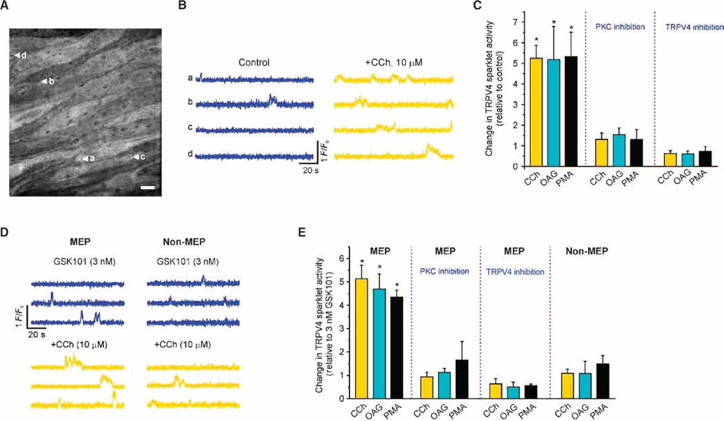
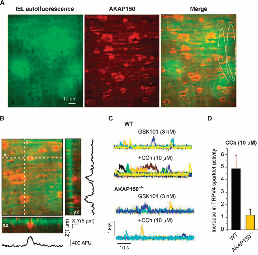
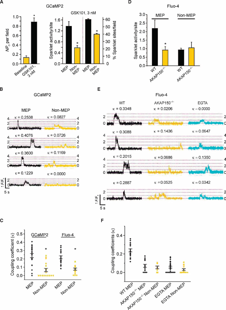
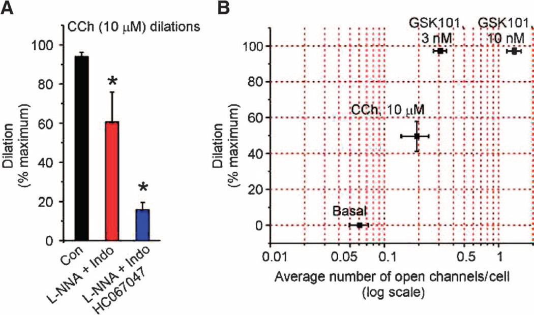
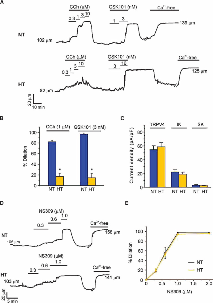
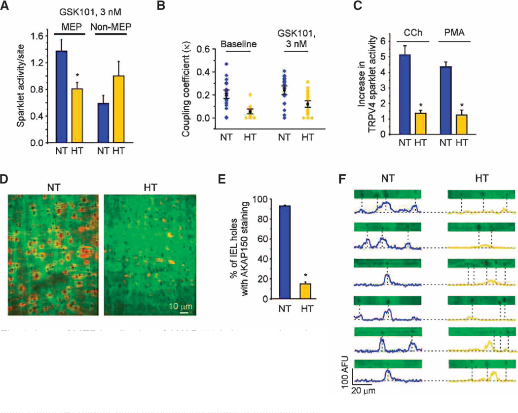
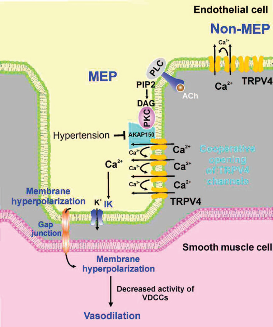
Endothelial cell dysfunction, characterized by a diminished response to endothelial cell-dependent vasodilators, is a hallmark of hypertension. TRPV4 channels play a major role in endothelial-dependent vasodilation, a function mediated by local Ca(2+) influx through clusters of functionally coupled TRPV4 channels rather than by a global increase in endothelial cell Ca(2+). We showed that stimulation of muscarinic acetylcholine receptors on endothelial cells of mouse arteries exclusively activated TRPV4 channels that were localized at myoendothelial projections (MEPs), specialized regions of endothelial cells that contact smooth muscle cells. Muscarinic receptor-mediated activation of TRPV4 depended on protein kinase C (PKC) and the PKC-anchoring protein AKAP150, which was concentrated at MEPs. Cooperative opening of clustered TRPV4 channels specifically amplified Ca(2+) influx at MEPs. Cooperativity of TRPV4 channels at non-MEP sites was much lower, and cooperativity at MEPs was greatly reduced by chelation of intracellular Ca(2+) or AKAP150 knockout, suggesting that Ca(2+) entering through adjacent channels underlies the AKAP150-dependent potentiation of TRPV4 activity. In a mouse model of angiotensin II-induced hypertension, MEP localization of AKAP150 was disrupted, muscarinic receptor stimulation did not activate TRPV4 channels, cooperativity among TRPV4 channels at MEPs was weaker, and vasodilation in response to muscarinic receptor stimulation was reduced. Thus, endothelial-dependent dilation of resistance arteries is enabled by MEP-localized AKAP150, which ensures the proximity of PKC to TRPV4 channels and the coupled channel gating necessary for efficient communication from endothelial to smooth muscle cells in arteries. Disruption of this molecular assembly may contribute to altered blood flow in hypertension.


Figures

Fig. 1
Fig. 1. CCh activates TRPV4 sparklets at MEPs through the PLC-DAG-PKC pathway
TRPV4 Ca2+ sparklets were recorded in en face preparations of mesenteric arteries from GCaMP2 mice in the presence of CPA (30 µM). (A) Grayscale image showing ECs in a field of view. The black holes in the internal elastic lamina correspond to sites where MEPs are located. The letter-labeled white arrows indicate four different TRPV4 sparklet sites at MEPs. (B) Representative F/F0 traces from the TRPV4 sparklet sites at MEPs indicated in (A) under basal conditions before (control) and after CCh addition. (C) Bar graph summarizing the effects of CCh (10 µM), OAG (10 µM), and PMA (10 nM) on TRPV4 sparklet activity at MEP sites in the presence or absence of the PKC inhibitor Gö-6976 (1 µM) or the TRPV4 inhibitor HC-067047 (1 µM). CPA-only was used as a control except in the PKC inhibition group, where CPA + Gö-6976 was used as a control. Data are means ± SEM (n= 5 to 10 fields). (D) Representative F/F0 traces from three different MEP and non-MEP sparklet sites before (blue) and after (yellow) CCh addition in the presence of 3 nM GSK101. (E) Bar graph summarizing the effects of CCh, OAG, and PMA on TRPV4 sparklets at MEP and non-MEP sites in the presence of GSK101 (3 nM) and in the presence or absence of Gö-6976 or HC-067047. Data are means ± SEM (n = 4 to 7 fields). Sparklet activity in the presence of CPA (30 µM) and GSK101 (3 nM) was used as a control, except for the PKC inhibition group, where sparklet activity in the presence of CPA, GSK101, and Gö-6976 was used as a control. In (C) and (E), Gö-6976 was added 10 min before CCh, OAG, or PMA treatment, whereas HC-067047 was added after CCh, OAG, or PMA (for 10 min). *P < 0.05 for columns 1 to 3 versus control and versus columns 7 to 9 [one-way analysis of variance (ANOVA) with post hoc Bonferroni test].
Fig. 2
Fig. 2. MEP-localized AKAP150 is required for CCh-induced activation of TRPV4 sparklets
(A) Images of an en face mesenteric artery preparation from a C57BL6 mouse showing AKAP150 immunostaining (center, red) at the level of the inner elastic lamina (IEL) (left, green); 93.1 ± 0.8% of holes in the IEL exhibited AKAP150 immunostaining (n = 10 fields, five arteries). Dotted lines in the merged image indicate the outlines of two ECs. (B) Three-dimensional view along the z axis (3 µm, 0.1-µm optical slices) showing densities of AKAP150 fluorescence projecting through the depth of the IEL. Reconstructed xz and yz images show AKAP150 immunostaining along the MEP, with strongest staining toward the end of the projections. Traces are plot profiles for AKAP150 staining along x and y axes for 5-µm-wide transects through the projections shown in the three-dimensional view. (C) Representative F/F0 traces of TRPV4 Ca2+ sparklets in mesenteric arteries from wild-type (WT) mice (top) and AKAP150−/− mice (bottom). The experiments were performed in Fluo-4–loaded mesenteric arteries from WT and AKAP150−/− mice in the presence of CPA (30 µM) and GSK101. Each color represents a trace from an individual region of interest (ROI). (D) Bar plots illustrating the CCh-induced increase in sparklet activity in the ECs of mesenteric arteries from WT and AKAP150−/− mice in the presence of GSK101. Data are means ± SEM (n = 5 to 6 fields, four to five arteries; P < 0.01, t test). Information on the number and size of holes, and the depth of the IEL is provided in table S4.
Fig. 3
Fig. 3. AKAP150 and local Ca2+ are responsible for coupled gating of TRPV4 channels at MEPs
TRPV4 Ca2+ sparklets were recorded in mesenteric arteries from GCaMP2 mice or Fluo-4–loaded mesen-teric arteries in en face preparations in the presence of CPA (30 µM) and GSK101 (3 nM). (A) Left: Effect of GSK101 on the average number of active TRPV4 channels (NPO) per field (n = 7 fields, 5 arteries). Right: Sparklet activity per site (*P < 0.05, t test; n = 21 to 38 sites) and the percentage of sparklet sites per field at MEP and non-MEP sites (*P< 0.05, t test; n=7 fields). (B) Representative F/F0 traces from four different TRPV4 Ca2+ sparklet sites at MEP and non-MEP locations on the EC membrane [quantal level: 0.19 ΔF/F0 (1)]. Respective coupling coefficient (κ) values are shown above each trace. (C) Scatter plot indicating individual κ values for sparklet sites at MEP and non-MEP locations. The mean coupling coefficient and SEM for each group are indicated (P < 0.01, t test). (D) Sparklet activity per site at MEP and non-MEP locations in mesenteric arteries from WT and AKAP150−/− mice (*P < 0.05, t test; 14 to 25 sites). (E) Representative F/F0 traces from four different TRPV4 Ca2+ sparklet sites at MEPs from WT mice in the absence (left, black) or presence (right, cyan) of EGTA-AM (10 µM for 10 min at 36°C) or AKAP150−/− (middle, yellow) mice. Respective κ values are shown above each F/F0 record [quantal level: 0.29 ΔF/F0 (1)]. (F) Scatter plot showing individual κ values for sparklet sites at MEPs from the groups in (E). The plot also shows the mean coupling coefficient and SEM for each group (n = 8 to 27 sites). A one-way ANOVA with post hoc Bonferroni test was used to estimate P values. P < 0.05 for WT MEP versus all other groups, and for WT non-MEP versus EGTA non-MEP.
Fig. 4
Fig. 4. Activation of less than one TRPV4 channel per EC causes maximum dilation of mesenteric arteries
(A) Bar graph showing dilatory responses to CCh alone (Con) and in the presence of the NOS and COX inhibitors l-NNA (100 µM) and indomethacin (Indo; 10 µM), respectively, to isolate the EDH component of the dilation, or in the presence of l-NNA, Indo, and HC-067047 (1 µM; TRPV4 inhibitor), to identify the EDH-mediated dilation that is dependent on TRPV4. Maximal dilation was defined as that in the presence of Ca2+-free PSS. Data are means ± SEM (n = 5 to 7 arteries); *P < 0.05 using one-way ANOVA with post hoc Bonferroni test. (B) HC-067047-sensitive (TRPV4) component of dilation in response to CCh, and GSK101 was plotted as a function of average TRPV4 channel activity per EC. The average number of TRPV4 channels activated per EC throughout the recording duration of 2 min was estimated by dividing the fluorescence integrals from all sparklet sites in an EC by the fluorescence integral of one quantal level (one channel) for 2 min. Data are means ± SEM (n = 5 to 7 arteries for dilation; 14 to 26 cells for average number of channels per cell).
Fig. 5
Fig. 5. EDH-mediated dilation is absent in arteries from hypertensive mice, but the function of individual ion channels is unaltered
Dilation studies in pressurized (80 mmHg) third-order mesenteric arteries from normotensive (NT) and hypertensive (HT) mice. (A) Representative vessel internal diameter traces in response to CCh or GSK101. (B) Summary of diameter data comparing the responses of NT and HT mouse arteries to CCh and GSK101 (n = 5 to 6 arteries from five NT and HT mice; *P < 0.01, t test). Data are means ± SEM. The maximum vessel dilation was determined from the diameter obtained in Ca2+-free solution. (C) Summary data showing maximum current densities of TRPV4 (at +100 mV), IK, and SK channels (both at 0 mV) in ECs from NT and HT mice (n= 5 to 8 cells). (D and E) Representative diameter traces (D) and summary of diameter data (E) showing dilation in response to the IK and SK channel opener NS309 (0.3 to 2 µM) in mesenteric arteries from NT and HT mice. Data are means ± SEM (n = 5 to 12 arteries). The averaged values for internal diameters are provided in tables S2 to S4.
Fig. 6
Fig. 6. Loss of MEP localization of AKAP150 in hypertensive mice leads to defective local Ca2+ signaling at MEPs and the loss of EDH-mediated dilation
(A) Sparklet activity per site for MEP and non-MEP sites in mesenteric arteries from NT and HT mice in the presence of GSK101 (10 to 13 arteries, n = 21 to 46 sites; *P < 0.05, t test). (B) Scatter plots summarizing coupling coefficient (κ) data for TRPV4 sparklets at MEPs in NT and HT mice (P < 0.01 for comparisons at baseline and in the presence of GSK101, t test). (C) Summary data illustrating the loss of activation of TRPV4 sparklets by CCh and PMA in HT arteries in the presence of GSK101 (3 nM; n = 4 to 5 arteries; *P < 0.01, t test). (D) Representative images of AKAP150 staining in en face preparations of mesenteric arteries from NT and HT mice. (E) Summary data of AKAP150-immunostaining data shown in (D). The number of holes with identifiable AKAP150 staining is expressed as a percentage of the total number of holes in the internal elastic lamina per field (n = 5 arteries; *P < 0.01, t test). (F) Plot profiles of AKAP150 immunostaining for representative horizontal transects (5 µm wide by 70 µm long) through the images in (D) encompassing holes. Dotted lines indicate the position of MEPs, located at holes in the IEL. Information on the number and sizes of holes, and the depth of the IEL in NT and HT mice is provided in table S4.
Fig. 7
Fig. 7. Proposed scheme for the organization and function of TRPV4 channels at MEPs
TRPV4 channels on the EC membrane exist in clusters of four channels at both MEP and non-MEP sites. Cooperative opening of TRPV4 channels depends on intracellular Ca2+ and AKAP150, which is concentrated at MEPs. Muscarinic receptor activation of TRPV4 channels occurs only at MEPs through AKAP150-anchored PKC. Ca2+ influx through activated TRPV4 channels activates IK channels and hyperpolarizes ECs, leading to hyperpolarization of SMC through gap junctions. Hyper-polarization reduces the activity of VDCCs in the SMCs, causing vaso-dilation. Endothelial dysfunction in Ang II–induced hypertension involves a loss of AKAP150 at MEPs, which uncouples muscarinic receptor signals to TRPV4 channels and disrupts cooperativity among TRPV4 channels. Although the current consensus is that EDH primarily involves electronic spread to the SMCs through gap junctions, soluble endothelium-derived hyperpolarizing factors (EDHFs, not shown) are released as a result of IK and SK channel activation may also contribute in certain circumstances.

Similar articles

Cited by

References

    1. Furchgott RF, Zawadzki JV. The obligatory role of endothelial cells in the relaxation of arterial smooth muscle by acetylcholine. Nature. 1980;288:373–376. - PubMed
    1. Dora KA. Coordination of vasomotor responses by the endothelium. Circ. J. 2010;74:226–232. - PubMed
    1. Fukao M, Hattori Y, Kanno M, Sakuma I, Kitabatake A. Sources of Ca2+ in relation to generation of acetylcholine-induced endothelium-dependent hyperpolarization in rat mesenteric artery. Br. J. Pharmacol. 1997;120:1328–1334. - PMC - PubMed
    1. Ledoux J, Taylor MS, Bonev AD, Hannah RM, Solodushko V, Shui B, Tallini Y, Kotlikoff MI, Nelson MT. Functional architecture of inositol 1,4,5-trisphosphate signaling in restricted spaces of myoendothelial projections. Proc. Natl. Acad. Sci. U.S.A. 2008;105:9627–9632. - PMC - PubMed
    1. Sandow SL, Senadheera S, Grayson TH, Welsh DG, Murphy TV. Calcium and endothelium-mediated vasodilator signaling. Adv. Exp. Med. Biol. 2012;740:811–831. - PubMed

Publication types

MeSH terms