Review
. 2014 Jul;141(14):2750-9.
doi: 10.1242/dev.104794.

Cellular and physical mechanisms of branching morphogenesis

Affiliations
Review

Cellular and physical mechanisms of branching morphogenesis

Victor D Varner et al. Development. 2014 Jul.

Abstract

Branching morphogenesis is the developmental program that builds the ramified epithelial trees of various organs, including the airways of the lung, the collecting ducts of the kidney, and the ducts of the mammary and salivary glands. Even though the final geometries of epithelial trees are distinct, the molecular signaling pathways that control branching morphogenesis appear to be conserved across organs and species. However, despite this molecular homology, recent advances in cell lineage analysis and real-time imaging have uncovered surprising differences in the mechanisms that build these diverse tissues. Here, we review these studies and discuss the cellular and physical mechanisms that can contribute to branching morphogenesis.

Keywords: Bifurcation; Mechanical stress; Pattern; Proliferation; Tension.


Figures

Fig. 1.
Fig. 1.
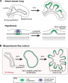
Branching via patterned or differential cell proliferation. Differential rates of cell proliferation have been hypothesized to induce branching in a number of developing organs, including the lung, kidney and salivary gland. In each of these cases, elevated levels of proliferation have been observed in nascent epithelial buds. (A) As a representative example, the developing mouse lung is shown. Given the observed patterns of proliferation, it is generally thought that growth factor expression (blue) in the neighboring mesenchyme stimulates localized growth/cell proliferation (green) in the epithelium, which initiates the formation of new epithelial branches. (B) Lung epithelial explants, denuded of mesenchyme and embedded in 3D gels of reconstituted basement membrane protein, have also been shown to branch in culture. Here again, elevated proliferation was observed in incipient branches, but only after these branches had already formed. No proliferative pre-pattern was observed in the epithelium prior to branching.
Fig. 2.
Fig. 2.
Branching via invasion. Collective cell migration drives branching morphogenesis in the Drosophila trachea and vertebrate vasculature. (A) In the Drosophila trachea, the expression of the FGF homolog Branchless (Bnl, green) specifies the tip cell, which inhibits tip cell phenotype in the neighboring stalk cells through Delta-Notch signaling. The collective migrates toward the source of Bnl. (B) This collective migration is mediated by at least two types of mechanical force. The tip cell induces tensile forces (red arrows) on the surrounding tissue, and pulls the stalk cells forward. At the same time, the stalk cells intercalate with each other, and these cellular rearrangements generate sufficient pushing forces (blue arrows) to move the collective forward.
Fig. 3.
Fig. 3.
Branching via epithelial folding. The collective folding of cells in an epithelium can produce new branches. Changes in epithelial shape can occur by: (A) apical constriction of cells within a localized region (pink) of the epithelium; (B) differential growth, caused by increased cell division in the epithelium (pink) relative to an adjacent tissue (white); or (C) mechanical buckling, which casts the epithelium into a wave-like morphology when loaded with sufficient compressive force, possibly produced by adjacent tissues.
Fig. 4.
Fig. 4.
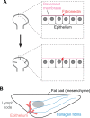
Matrix-mediated branching. The ECM plays an instructive role in branching morphogenesis in some organs. (A) In the salivary gland, fibrils of fibronectin (red) are deposited at the sites of new clefts (gray) in the epithelium. (B) In the mammary gland, fibrils of collagen (blue) present within the stroma may act as guidance cues for the elongation of nascent epithelial branches (red) during morphogenesis.

Similar articles

Cited by

References

    1. Affolter M., Caussinus E. (2008). Tracheal branching morphogenesis in Drosophila: new insights into cell behaviour and organ architecture. Development 135, 2055-2064. 10.1242/dev.014498 - DOI - PubMed
    1. Alcaraz J., Mori H., Ghajar C. M., Brownfield D., Galgoczy R., Bissell M. J. (2011). Collective epithelial cell invasion overcomes mechanical barriers of collagenous extracellular matrix by a narrow tube-like geometry and MMP14-dependent local softening. Integr. Biol. (Camb.) 3, 1153-1166. 10.1039/c1ib00073j - DOI - PMC - PubMed
    1. Alescio T., Cassini A. (1962). Induction in vitro of tracheal buds by pulmonary mesenchyme grafted on tracheal epithelium. J. Exp. Zool. 150, 83-94. 10.1002/jez.1401500202 - DOI - PubMed
    1. Alescio T., Colombo Piperno E. (1967). A quantitative assessment of mesenchymal contribution to epithelial growth rate in mouse embryonic lung developing in vitro. J. Embryol. Exp. Morphol. 17, 213-227 - PubMed
    1. Alescio T., Di Michele M. (1968). Relationship of epithelial growth to mitotic rate in mouse embryonic lung developing in vitro. J. Embryol. Exp. Morphol. 19, 227-237 - PubMed

Publication types