. 2014 Jul 9:4:5635.
doi: 10.1038/srep05635.

Generation of muscular dystrophy model rats with a CRISPR/Cas system

Affiliations

Generation of muscular dystrophy model rats with a CRISPR/Cas system

Katsuyuki Nakamura et al. Sci Rep. .

Abstract

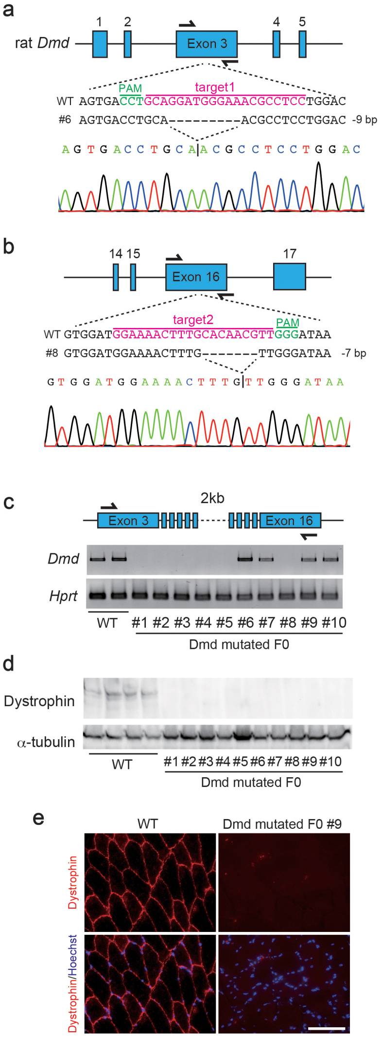
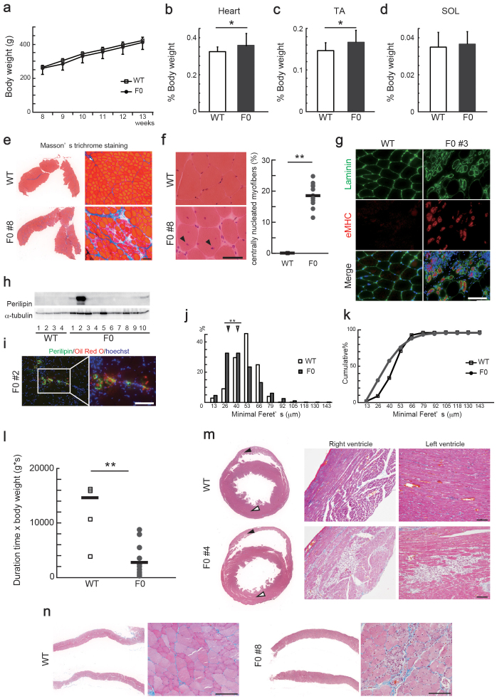
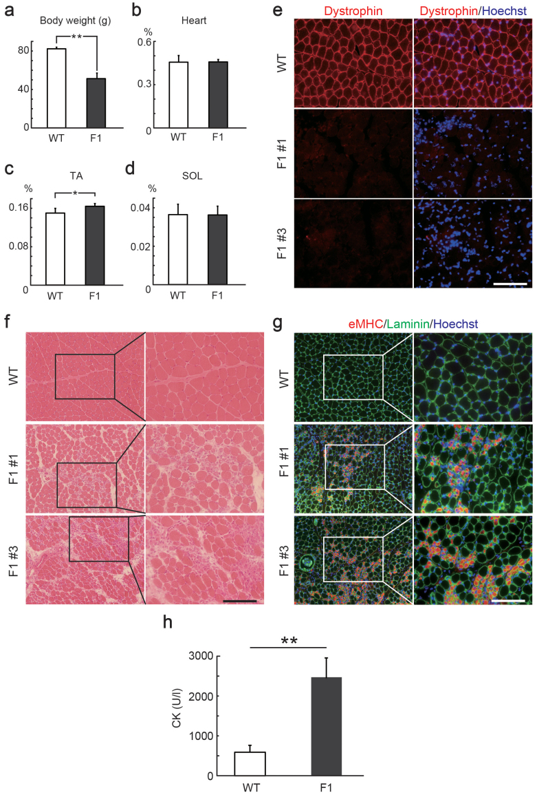
Duchenne muscular dystrophy (DMD) is an X-linked lethal muscle disorder caused by mutations in the Dmd gene encoding Dystrophin. DMD model animals, such as mdx mice and canine X-linked muscular dystrophy dogs, have been widely utilized in the development of a treatment for DMD. Here, we demonstrate the generation of Dmd-mutated rats using a clustered interspaced short palindromic repeats (CRISPR)/Cas system, an RNA-based genome engineering technique that is also adaptive to rats. We simultaneously targeted two exons in the rat Dmd gene, which resulted in the absence of Dystrophin expression in the F0 generation. Dmd-mutated rats exhibited a decline in muscle strength, and the emergence of degenerative/regenerative phenotypes in the skeletal muscle, heart, and diaphragm. These mutations were heritable by the next generation, and F1 male rats exhibited similar phenotypes in their skeletal muscles. These model rats should prove to be useful for developing therapeutic methods to treat DMD.


Figures

Figure 1
Figure 1. Generation of Dmd-mutated rats via the CRISPR/Cas9 system.
Scheme for targeting the rat Dmd gene at exons 3 (a) and 16 (b) using CRISPR/Cas9. The gRNA sequences (magenta) and protospacer adjacent motif (PAM; green) are labeled. Representative sequences and their waveform data are presented below. (c) RT-PCR for Dmd and Hprt in wild type (WT) and Dmd-mutated F0 rats. These images were cropped and full-length gels are shown in Supplementary Figure 8a and b. (d) Immunoblotting for Dystrophin and α-tubulin in tibialis anterior (TA) muscles of WT and Dmd-mutated F0 rats. These images were cropped and full-length blots are shown in Supplementary Figure 8c and d. (e) Immunostaining for Dmd in WT and Dmd-mutated F0 rats. Scale bar = 100 μm.
Figure 2
Figure 2. Pathological analyses of Dmd-mutated F0 rats.
(a) Body weight and weights of the (b) heart, (c) tibialis anterior (TA), and (d) soleus (SOL) muscles relative to body weight of Dmd-mutated F0 (n = 10) and age-matched wild type (WT) rats (n = 4). *P < 0.05. (e) Masson's trichrome staining of TA muscles in 13-week-old WT and Dmd-mutated F0 rats. Scale bar = 100 μm. (f) Hematoxylin & eosin (H&E) staining of TA muscles in 13-week-old WT and Dmd-mutated F0 rats. Scale bar = 50 μm. Myofibers with central nuclei were indicated with arrowheads and quantified in the right plots (WT; n = 4, Dmd-mutated F0; n = 10). Bars represent the mean value of each group. **P < 0.01. (g) Cross sections of TA muscles in 13-week-old WT and Dmd-mutated F0 rats stained with anti-laminin (green) and anti-embryonic myosin heavy chain (eMHC, red) antibody. Scale bar = 100 μm. (h) Immunoblotting for Perilipin and α-tubulin in TA muscles of WT and Dmd-mutated rats. Full-length blots are shown in Supplementary Figure 8e and f. (i) Cross sections of TA muscles in 13-week-old WT and Dmd-mutated F0 rats stained with anti-Perilipin antibody (green) and Oil Red O (red). Scale bar = 100 μm. Relative distributions (j) and cumulative plots (k) of myofiber size in TA muscles of 13-week-old WT and F0 rats (WT, 2448 fibers and F0, 6612 fibers). Arrowheads indicate median values. **P < 0.01. (l) Results of wire-hang test in WT (n = 4) and Dmd-mutated F0 (n = 10) rats. Bars represent the mean value of each group. **P < 0.01. Masson's trichrome stainings of (m) hearts and (n) diaphragms of 13-week-old WT and Dmd-mutated F0 rats. Scale bar = 100 μm. Black and white arrowheads indicate right and left ventricles, respectively. Scale bar = 100 μm.
Figure 3
Figure 3. Inheritance of mutations in the rat Dmd gene generated by CRISPR/Cas9.
Comparison of body weight (a), weights of the heart (b), tibialis anterior (TA) muscle (c), and solaris (SOL) muscle (d) relative to body weight of 4-week-old wild type (WT) and F1 male rats. *P < 0.05, **P < 0.01. (e) Cross sections of TA muscles in 4-week-old WT and F1 male rats stained with anti-Dystrophin (red) antibody. Scale bar = 100 μm. (f) Hematoxylin and eosin (H&E) staining of TA muscles in 4-week-old WT and F1 male rats. Scale bar = 100 μm. (g) Cross sections of TA muscles in 4-week-old WT and F1 male rats stained with anti-laminin (green) and anti-embryonic myosin heavy chain (eMHC, red) antibody. Scale bar = 100 μm. (h) Creatine kinase (CK) activity of Dmd-mutated F1 (n = 5) and age-matched WT (n = 3) rats. **P < 0.01.

References

    1. Koenig M., Monaco A. P. & Kunkel L. M. The complete sequence of dystrophin predicts a rod-shaped cytoskeletal protein. Cell 53, 219–228 (1988). - PubMed
    1. Hoffman E. P., Brown R. H. Jr & Kunkel L. M. Dystrophin: the protein product of the Duchenne muscular dystrophy locus. Cell 51, 919–28 (1987). - PubMed
    1. Vainzof M. et al. Animal models for genetic neuromuscular diseases. J. Mol. Neurosci. 34, 241–248 (2008). - PubMed
    1. Kinali M. et al. Muscle histology vs MRI in Duchenne muscular dystrophy. Neurology 76, 346–353 (2011). - PMC - PubMed
    1. Sekiguchi M. et al.. A deficit of brain dystrophin impairs specific amygdala GABAergic transmission and enhances defensive behaviour in mice. Brain 132, 124–135 (2009). - PubMed

Publication types