Use of diffusion tensor imaging to assess the impact of normobaric hyperoxia within at-risk pericontusional tissue after traumatic brain injury
- PMID: 25005875
- PMCID: PMC4269721
- DOI: 10.1038/jcbfm.2014.123
Use of diffusion tensor imaging to assess the impact of normobaric hyperoxia within at-risk pericontusional tissue after traumatic brain injury
Abstract
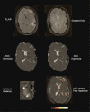
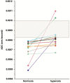
Ischemia and metabolic dysfunction remain important causes of neuronal loss after head injury, and we have shown that normobaric hyperoxia may rescue such metabolic compromise. This study examines the impact of hyperoxia within injured brain using diffusion tensor imaging (DTI). Fourteen patients underwent DTI at baseline and after 1 hour of 80% oxygen. Using the apparent diffusion coefficient (ADC) we assessed the impact of hyperoxia within contusions and a 1 cm border zone of normal appearing pericontusion, and within a rim of perilesional reduced ADC consistent with cytotoxic edema and metabolic compromise. Seven healthy volunteers underwent imaging at 21%, 60%, and 100% oxygen. In volunteers there was no ADC change with hyperoxia, and contusion and pericontusion ADC values were higher than volunteers (P<0.01). There was no ADC change after hyperoxia within contusion, but an increase within pericontusion (P<0.05). We identified a rim of perilesional cytotoxic edema in 13 patients, and hyperoxia resulted in an ADC increase towards normal (P=0.02). We demonstrate that hyperoxia may result in benefit within the perilesional rim of cytotoxic edema. Future studies should address whether a longer period of hyperoxia has a favorable impact on the evolution of tissue injury.
Figures


Similar articles
-
Normobaric hyperoxia does not improve derangements in diffusion tensor imaging found distant from visible contusions following acute traumatic brain injury.Sci Rep. 2017 Sep 29;7(1):12419. doi: 10.1038/s41598-017-12590-2. Sci Rep. 2017. PMID: 28963497 Free PMC article.
-
Microstructural basis of contusion expansion in traumatic brain injury: insights from diffusion tensor imaging.J Cereb Blood Flow Metab. 2013 Jun;33(6):855-62. doi: 10.1038/jcbfm.2013.11. Epub 2013 Feb 20. J Cereb Blood Flow Metab. 2013. PMID: 23423189 Free PMC article.
-
Use of diffusion tensor imaging to assess the vasogenic edema in traumatic pericontusional tissue.Neurocirugia (Engl Ed). 2021 Jul-Aug;32(4):161-169. doi: 10.1016/j.neucie.2020.05.001. Neurocirugia (Engl Ed). 2021. PMID: 34218876
-
[Normobaric hyperoxia therapy for patients with traumatic brain injury].Ann Fr Anesth Reanim. 2012 Mar;31(3):224-7. doi: 10.1016/j.annfar.2011.11.009. Epub 2012 Feb 3. Ann Fr Anesth Reanim. 2012. PMID: 22305404 Review. French.
-
Normobaric hyperoxia therapy for traumatic brain injury and stroke: a review.Br J Neurosurg. 2009 Dec;23(6):576-84. doi: 10.3109/02688690903050352. Br J Neurosurg. 2009. PMID: 19922270 Review.
Cited by
-
Intensive Care in Traumatic Brain Injury Including Multi-Modal Monitoring and Neuroprotection.Med Sci (Basel). 2019 Feb 26;7(3):37. doi: 10.3390/medsci7030037. Med Sci (Basel). 2019. PMID: 30813644 Free PMC article. Review.
-
Neuroanatomical Substrates and Symptoms Associated With Magnetic Resonance Imaging of Patients With Mild Traumatic Brain Injury.JAMA Netw Open. 2021 Mar 1;4(3):e210994. doi: 10.1001/jamanetworkopen.2021.0994. JAMA Netw Open. 2021. PMID: 33734414 Free PMC article.
-
Neuroprotection strategies in traumatic brain injury: Studying the effectiveness of different clinical approaches.Surg Neurol Int. 2024 Jan 26;15:29. doi: 10.25259/SNI_773_2023. eCollection 2024. Surg Neurol Int. 2024. PMID: 38344087 Free PMC article. Review.
-
Hyperoxia in intensive care, emergency, and peri-operative medicine: Dr. Jekyll or Mr. Hyde? A 2015 update.Ann Intensive Care. 2015 Dec;5(1):42. doi: 10.1186/s13613-015-0084-6. Epub 2015 Nov 19. Ann Intensive Care. 2015. PMID: 26585328 Free PMC article.
-
Dangers of hyperoxia.Crit Care. 2021 Dec 19;25(1):440. doi: 10.1186/s13054-021-03815-y. Crit Care. 2021. PMID: 34924022 Free PMC article. Review.
References
-
- Coles JP, Fryer TD, Smielewski P, Rice K, Clark JC, Pickard JD, et al. Defining ischemic burden after traumatic brain injury using 15O PET imaging of cerebral physiology. J Cereb Blood Flow Metab. 2004;24:191–201. - PubMed
-
- Nortje J, Coles JP, Timofeev I, Fryer TD, Aigbirhio FI, Smielewski P, et al. Effect of hyperoxia on regional oxygenation and metabolism after severe traumatic brain injury: preliminary findings. Crit Care Med. 2008;36:273–281. - PubMed
-
- Menon DK, Coles JP, Gupta AK, Fryer TD, Smielewski P, Chatfield DA, et al. Diffusion limited oxygen delivery following head injury. Crit Care Med. 2004;32:1384–1390. - PubMed
-
- Ahn ES, Robertson CL, Vereczki V, Hoffman GE, Fiskum G. Normoxic ventilatory resuscitation following controlled cortical impact reduces peroxynitrite-mediated protein nitration in the hippocampus. J Neurosurg. 2008;108:124–131. - PubMed
Publication types
MeSH terms
Substances
Grants and funding
LinkOut - more resources
Full Text Sources
Other Literature Sources
Medical

