. 2014 Sep 11;124(11):1748-64.
doi: 10.1182/blood-2014-03-562710. Epub 2014 Jul 8.

Resolution of acute inflammation bridges the gap between innate and adaptive immunity

Affiliations

Resolution of acute inflammation bridges the gap between innate and adaptive immunity

Justine Newson et al. Blood. .

Abstract

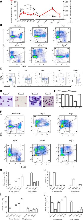
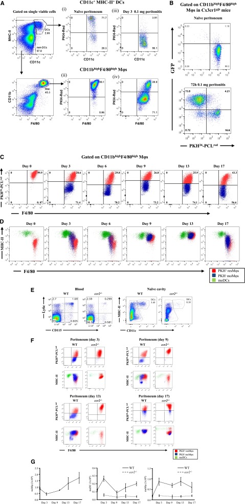
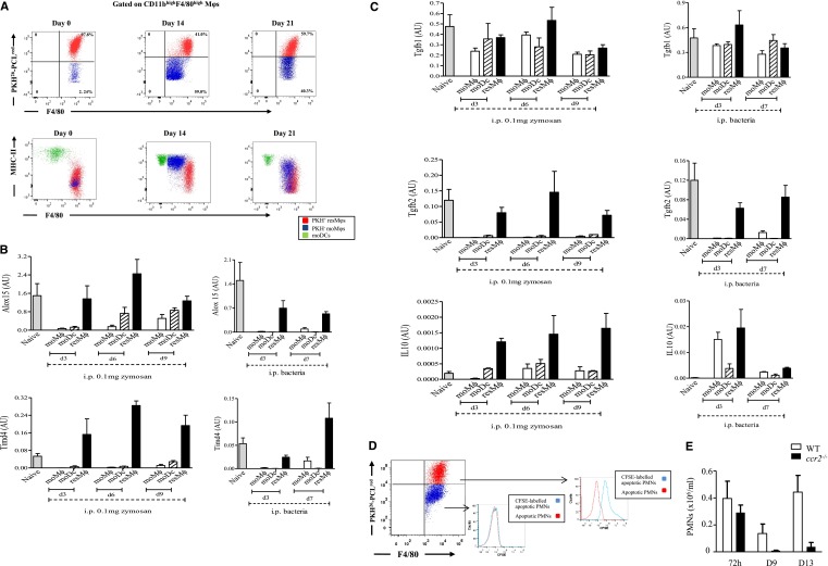
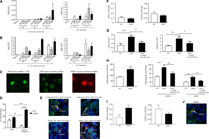
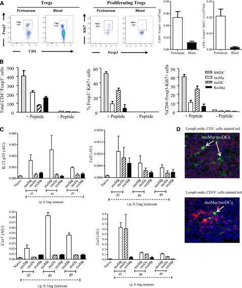
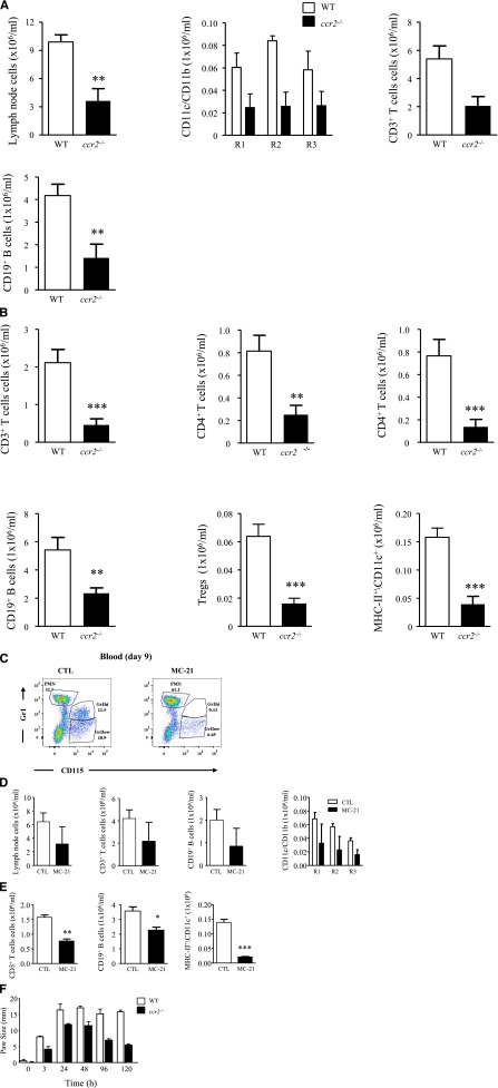
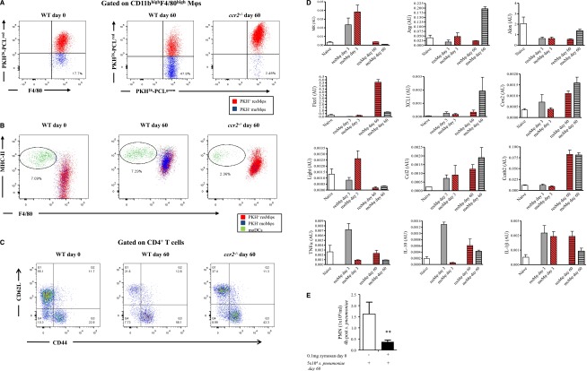
Acute inflammation is traditionally characterized by polymorphonuclear leukocytes (PMN) influx followed by phagocytosing macrophage (Mφs) that clear injurious stimuli leading to resolution and tissue homeostasis. However, using the peritoneal cavity, we found that although innate immune-mediated responses to low-dose zymosan or bacteria resolve within days, these stimuli, but not hyperinflammatory stimuli, trigger a previously overlooked second wave of leukocyte influx into tissues that persists for weeks. These cells comprise distinct populations of tissue-resident Mφs (resMφs), Ly6c(hi) monocyte-derived Mφs (moMφs), monocyte-derived dendritic cells (moDCs), and myeloid-derived suppressor cells (MDSCs). Postresolution mononuclear phagocytes were observed alongside lymph node expansion and increased numbers of blood and peritoneal memory T and B lymphocytes. The resMφs and moMφs triggered FoxP3 expression within CD4 cells, whereas moDCs drive T-cell proliferation. The resMφs preferentially clear apoptotic PMNs and migrate to lymph nodes to bring about their contraction in an inducible nitric oxide synthase-dependent manner. Finally, moMφs remain in tissues for months postresolution, alongside altered numbers of T cells collectively dictating the magnitude of subsequent acute inflammatory reactions. These data challenge the prevailing idea that resolution leads back to homeostasis and asserts that resolution acts as a bridge between innate and adaptive immunity, as well as tissue reprogramming.


Figures

Figure 1
Figure 1
Inflammation in response to low- vs high-dose zymosan in the mouse peritoneum. (A) Either 0.1 or 10 mg of zymosan was injected into the peritoneal cavity of separate groups of mice. (B) Polychromatic flow cytometry is shown carried out on inflammation driven by 0.1 mg of zymosan, highlighting monocyte/macrophage populations, whereas (C) depicts, among other cells types MDSCs, alongside their (D) histological appearance and ability to (E) suppress T-cell proliferation. (F) In contrast, flow cytometry is shown carried out on inflammation driven by a more aggressive dose of 10 mg of zymosan, highlighting monocyte/macrophage populations, whereas (G-H) summarizes the relative temporal profiles of monocyte/macrophages andPMNs in these 2 models. (I-J) Profiles of lymphocytes are shown. Data are presented as mean ± SEM for n = 8 mice/group. ***P < .005.
Figure 2
Figure 2
Temporal profiles of mononuclear phagocytes and DCs throughout inflammation, resolution, and postresolution/adaptive immunity phase. The gating strategy in supplemental Figure 4 was used to identify Mφ and DC populations in the peritoneal cavity of naïve mice. Using this approach, (A) the cell tracker dye PKH26-PCLred was injected into the cavity of mice and its labeling of tissue-resident DCs and resMφ determined in the naïve peritoneum (A, panels i-ii, respectively). These mice were then injected with 0.1 mg of zymosan, revealing (A, panel iii) the disappearance of DCs from the naïve peritoneum after inflammation and the presence of both (A, panel iv) PKH26-PCLred-positive resMφs and PKH26-PCLred-negative moMφs 72 hours post-zymosan. The origin of the latter as being Ly6chi-derived was confirmed using (B) CX3CRgfp mice. (C) The temporal and relative changes of resMφs vs moMφs (Ly6chi–monocyte-derived) from onset (4 hours), classic resolution (48-72 hours) and postresolution from day 6 onwards are shown. These data were further back-gated onto (D) MHC-II vs F4/80 to depict the overall temporal changes of mononuclear phagocytes and DCs throughout and after resolving inflammation. Further experiments were carried out using (E-F) ccr2−/− mice to prove the ly6chi origin of moMφs and MoDCs throughout resolution and postresolution with the (G) temporal profiles of resMφs, moMφs, and MoDCs shown. Data are presented as mean ± SEM for n = 6 mice/group.
Figure 3
Figure 3
Aspects of resolution-phase Mφ phenotype are conserved between sterile and infections resolving inflammation. (A) The temporal profile in resMφs, moMφs, and DCs are shown throughout the inflammatory and postresolution response to S pneumoniae. These cells, as well as the equivalent population from 0.1 mg of zymosan, were shown (B-C) FACSorted and subjected to reverse transcription polymerase chain reaction. Injecting CFSE-labeled apoptotic PMNs into the peritoneum 48 hours post 0.1 mg of zymosan confirmed that (D) resMφs preferentially phagocytosed apoptotic PMNs with a little role for moMφs in this process; data confirmed using (E) Ly6chi-deficinet ccr2−/− mice showing no buildup of PMNs in the cavity postresolution. Data are presented as mean ± SEM for n = 6 mice/group. i.p., intraperitoneal.
Figure 4
Figure 4
The resMφs bring about postresolution lymphocyte contraction in an iNOS-dependent manner. The resMφs from 0.1 mg of zymosan and S pneumonia-induced acute resolving inflammation revealed increased expression of (A) iNOS and (B) arginase. (C) Immunofluorescence was used to visualize the intracellular localization of iNOS in postresolution resMφs, whereas (D) confirmed their iNOS-dependent suppression of T-cell proliferation. (E) Some PKH26-PCLred-positive resMφs migrate to mesenteric lymph nodes and spleen day 9 post 0.1 mg zymosan. Migrated iNOS-expressing resMφs mediate immune suppression was illustrated in iNOS−/− mice, although the composition of the (F) naïve cavity is equivalent between knockouts and controls, and 14 days after inflammation lymphocyte numbers were greater in iNOS−/− mice (G) peritoneal cavity and (H) spleens with (I) effects in persisting in spleen for up to 6 weeks. (G-I) The reversal of adaptive immune responses in iNOS deficient animals by the intraperitoneal injection of PKH26-PCLred-positive resMφ from WT mice into iNOS−/− mice are shown. (J) Adoptively transferred resMφs (stained red) from WT mice migrated to the spleen of iNOS knockouts; CD3 cells are stained in green. The P value was ≤.05, as determined by ANOVA, followed by the Bonferroni t test or two-tailed Student t test, with data expressed as mean ± SEM for n = 6 mice/group. *P ≤ .05; **P < .01; ***P < .001.
Figure 5
Figure 5
Postresolution Mφs trigger FoxP3 expression in CD4 T cells. (A) The relative ratios of blood vs peritoneal Tregs was determined 9 days after zymosan injection (0.1 mg) with postresolution resM, moMφs, and moDCs incubated with CD4 T- and Trp-1 peptide (SGHNCGTCRPGWRGAACNQKILTVR) + TGF-β for 5 days and analyzed for the presence of (B) Foxp3 expression and effector T-cell proliferation. Analysis of these Mφ/DC populations revealed a (C) migratory phenotype that was (D) confirmed by injecting PKH26-PCLgreen into mice on day 6 post-zymosan (0.1 mg), which also had PKH26-PCLred injected into their naïve peritoneum to label resMφs. This resulted in infiltrating moMφs and moDCs labeling positively for only PKH26-PCLgreen (shown), whereas resMφs were labeled with both PKH26-PCLred and PKH26-PCLgreen (not shown). Data are presented as mean ± SEM for n = 6 mice/group.
Figure 6
Figure 6
Postresolution adaptive immunity is dampened in ccr2−/− mice and with therapeutic depletion of postresolution Ly6chi monocyte. Zymosan (0.1 mg) was injected into ccr2−/− mice after a determination of (A) mesenteric lymph node CD11c+ DCs (R1), CD11c+/CD11b+ Mφs (R2), and CD11b+ Mφs (R3), as well as CD3 and CD19 T and B cells on day 9. (B) The corresponding distribution of lymphocyte populations in the peritoneum at the same time point is shown. MC-21 was given to WT mice 3 days after zymosan, and its effect on (C) Ly6chi monocyte populations was determined in the blood of naive animals and (D) mesenteric lymph node lymphocyte and Mφs/DCs alongside (E) lymphocytes and Mφs/DCs in the peritoneum day 9 post-zymosan injection (0.1 mg). (F) Reduced inflammation in ccr2−/− mice bearing a delayed type hypersensitivity reaction is shown. The P value was ≤.05, as determined by ANOVA, followed by the Bonferroni t test or two-tailed Student t test, with data expressed as mean ± SEM for n = 6 mice/group. *P ≤ .05; **P < .01; ***P < .001.
Figure 7
Figure 7
A state of adapted homeostasis is experienced after resolving inflammation. PKH26-PCLred was injected into the cavity of naïve WT and ccr2−/− mice followed 2 hours later by 0.1 mg of zymosan. Six days after zymosan PKH26-PCLgreen was injected to distinguish resMφs (PKH26-PCLgred and PKH26-PCLgreen) from infiltrating moMφs/DCs (PKH26-PCLgreen only). The peritoneal cavity of these mice was examined 60 days after the initial zymosan injection revealing (A) a population of moMφs that were PKH26-PCLgreen, but were absent in ccr2−/− mice alongside (B) moDC numbers and (C) T-cell activation markers. (D) The resMφs and moDCs were FASCsorted for phenotypic analysis, whereas (E) the impact of postresolution altered homeostasis to a second hit of S pneumonia was determined on day 60. The P value was ≤.05, as determined by ANOVA, followed by the Bonferroni t test or two-tailed Student t test, with data expressed as mean ± SEM for n = 6 mice/group. *P ≤ .05; **P < .01; ***P < .001.

Comment in

References

    1. Serhan CN, Brain SD, Buckley CD, et al. Resolution of inflammation: state of the art, definitions and terms. FASEB J. 2007;21(2):325–332. - PMC - PubMed
    1. Serhan CN, Savill J. Resolution of inflammation: the beginning programs the end. Nat Immunol. 2005;6(12):1191–1197. - PubMed
    1. Jamieson T, Cook DN, Nibbs RJ, et al. The chemokine receptor D6 limits the inflammatory response in vivo. Nat Immunol. 2005;6(4):403–411. - PubMed
    1. Lohr J, Knoechel B, Abbas AK. Regulatory T cells in the periphery. Immunol Rev. 2006;212:149–162. - PubMed
    1. Trivedi SG, Newson J, Rajakariar R, et al. Essential role for hematopoietic prostaglandin D2 synthase in the control of delayed type hypersensitivity. Proc Natl Acad Sci USA. 2006;103(13):5179–5184. - PMC - PubMed

Publication types

MeSH terms