. 2014 Aug 22;289(34):23504-19.
doi: 10.1074/jbc.M114.553305. Epub 2014 Jul 8.

Identification of multifaceted binding modes for pyrin and ASC pyrin domains gives insights into pyrin inflammasome assembly

Affiliations

Identification of multifaceted binding modes for pyrin and ASC pyrin domains gives insights into pyrin inflammasome assembly

Parimala R Vajjhala et al. J Biol Chem. .

Abstract

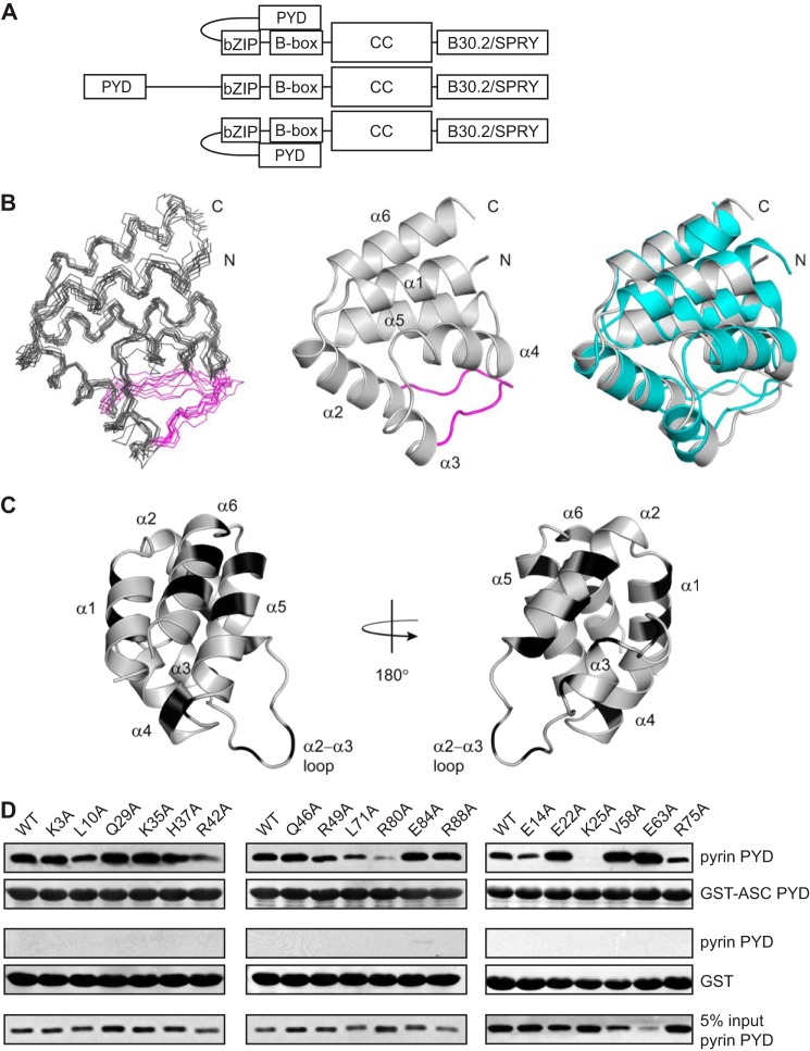
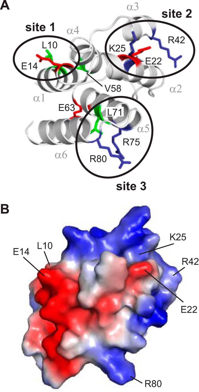
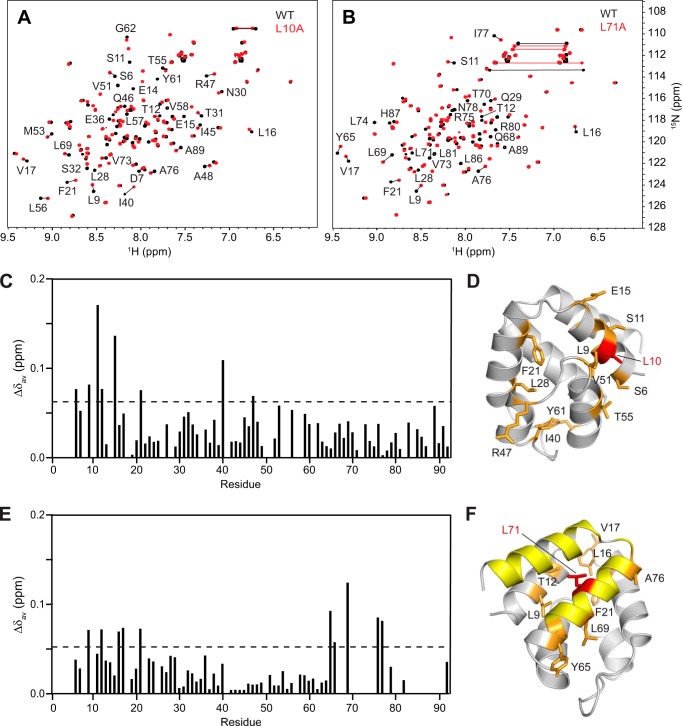
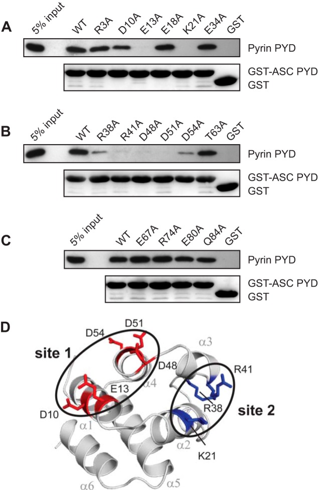
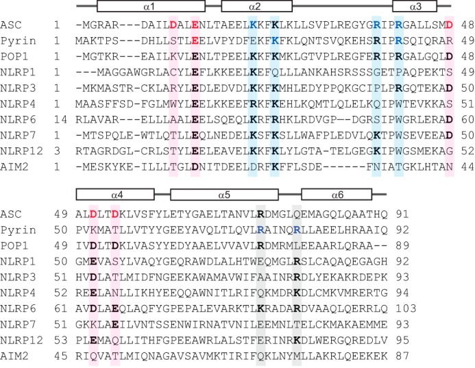
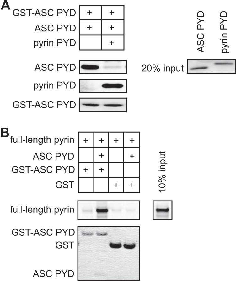
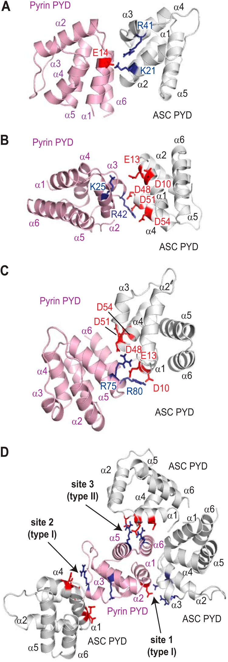
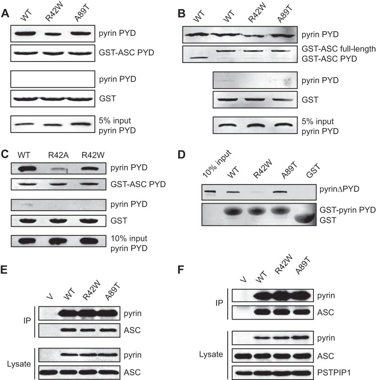
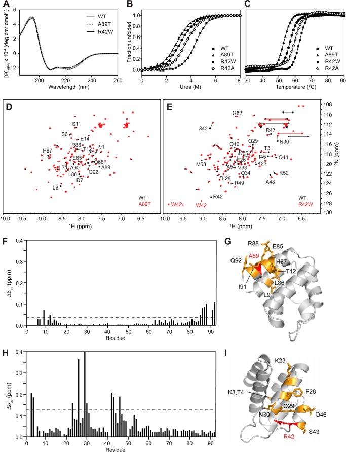
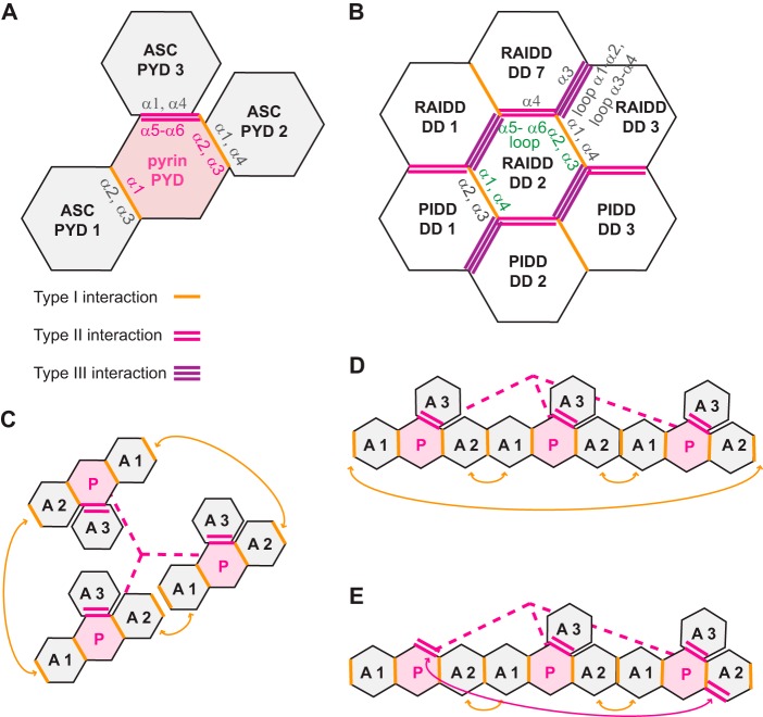
Inflammasomes are macromolecular complexes that mediate inflammatory and cell death responses to pathogens and cellular stress signals. Dysregulated inflammasome activation is associated with autoinflammatory syndromes and several common diseases. During inflammasome assembly, oligomerized cytosolic pattern recognition receptors recruit procaspase-1 and procaspase-8 via the adaptor protein ASC. Inflammasome assembly is mediated by pyrin domains (PYDs) and caspase recruitment domains, which are protein interaction domains of the death fold superfamily. However, the molecular details of their interactions are poorly understood. We have studied the interaction between ASC and pyrin PYDs that mediates ASC recruitment to the pyrin inflammasome, which is implicated in the pathogenesis of familial Mediterranean fever. We demonstrate that both the ASC and pyrin PYDs have multifaceted binding modes, involving three sites on pyrin PYD and two sites on ASC PYD. Molecular docking of pyrin-ASC PYD complexes showed that pyrin PYD can simultaneously interact with up to three ASC PYDs. Furthermore, ASC PYD can self-associate and interact with pyrin, consistent with previous reports that pyrin promotes ASC clustering to form a proinflammatory complex. Finally, the effects of familial Mediterranean fever-associated mutations, R42W and A89T, on structural and functional properties of pyrin PYD were investigated. The R42W mutation had a significant effect on structure and increased stability. Although the R42W mutant exhibited reduced interaction with ASC, it also bound less to the pyrin B-box domain responsible for autoinhibition and hence may be constitutively active. Our data give new insights into the binding modes of PYDs and inflammasome architecture.

Keywords: Death Domain; Familial Mediterranean Fever; Inflammasome; Inflammation; Innate Immunity; Mutagenesis; Pattern Recognition Receptor (PRR); Protein Complex; Protein-Protein Interaction; Pyrin.


Figures

FIGURE 1.
FIGURE 1.
Identification of residues on pyrin PYD that are important for interaction with ASC PYD. A, schematic representation of a pyrin trimer indicating positions of the PYD (residues 1–92), bZIP domain (residues 266–270), B-box domain (residues 375–407), coiled-coil domain (CC, residues 408–594), and B30.2/SPRY domain (residues 598–774). The central pyrin molecule is shown as a linear schematic, whereas the molecules on either side illustrate the intramolecular interaction between the PYD and B-box. B, from left to right, ensemble of 10 NMR-derived structures of pyrin PYD superimposed over the backbone atoms (N, Cα, and C′) of residues 5–92; ribbon diagram of the lowest energy pyrin PYD structure illustrating the six α-helices and the α2-α3 loop, which is colored magenta; and superposition of the pyrin PYD (gray) and ASC PYD (Protein Data Bank code 1UCP, cyan) (32) structures for backbone Cα residues 5–92 of pyrin PYD and residues 4–91 of ASC PYD. C, ribbon representation of the pyrin PYD structure with residues mutated in this study shown in black. The residues mutated include Lys3, Leu10, and Glu14 (helix α1); Glu22, Lys25, and Gln29 (helix α2); Lys35 and His37 (α2-α3 loop); Arg42 and Gln46 (helix α3); Arg49 (α3-α4 loop); Val58 (helix α4); Glu63, Leu71, and Arg75 (helix α5); and Arg80, Glu84, and Arg88 (helix α6). D, purified His6-tagged WT and mutant pyrin PYDs were used for in vitro binding assays with purified bead-bound GST-ASC PYD or GST alone. Bound protein was eluted with SDS-PAGE sample buffer, subjected to SDS-PAGE, and then transferred to a PVDF membrane. The bound His6-tagged WT and mutant pyrin PYDs detected by immunoblotting with an anti-His antibody are shown above a Ponceau S stain from the same blot to detect GST-ASC PYD or GST alone. An amount representing 5% of the input of WT or mutant pyrin PYDs used for binding studies is also shown.
FIGURE 2.
FIGURE 2.
Residues important for interaction of pyrin PYD with ASC PYD localize to three clusters. A, ribbon representation of the pyrin PYD structure highlighting residues that affect interaction when mutated. Basic, acidic, and hydrophobic residues are colored blue, red, and green, respectively. Clusters of residues that form an interaction site are circled. B, surface electrostatic potential of pyrin PYD shown in the same orientation as in A and indicating residues important for interaction.
FIGURE 3.
FIGURE 3.
The majority of pyrin PYD mutant proteins retain structural integrity. A–F, overlay of the two-dimensional 1H-15N HSQC spectra of wild-type (black) and mutant (red) pyrin PYD proteins E14A, E22A, R42A, E63A, R75A, and R80A, respectively. All spectra were recorded in 50 mm sodium phosphate, pH 4, and 150 mm NaCl at 25 °C. Residues that show chemical shift changes are indicated with the one-letter amino acid code and sequence number. Horizontal lines connect peaks corresponding to side chain NH2 groups of Asn and Gln residues that exhibit changes in chemical shifts. G–L, ribbon diagrams of the pyrin PYD structure showing residues with their chemical shifts perturbed by the E14A, E22A, R42A, E63A, R75A, and R80A mutations, respectively. Mutated residues are colored red, and residues with chemical shift changes are colored orange, and their side chains are shown.
FIGURE 4.
FIGURE 4.
Pyrin PYD mutations L10A and L71A perturb the protein structure. A and B, overlay of the two-dimensional 1H-15N HSQC spectra of wild-type (black) and mutant (red) pyrin PYD proteins L10A and L71A, respectively. All spectra were recorded in 50 mm sodium phosphate, pH 4, and 150 mm NaCl at 25 °C. Residues that show chemical shift changes are indicated with the one-letter amino acid code and sequence number. Horizontal lines connect peaks corresponding to side chain NH2 groups of Asn and Gln residues that exhibit changes in chemical shifts. C and E, histograms of the weighted backbone amide chemical shift changes (Δδav) versus residue number for the L10A and L71A mutants, respectively. The dashed line represents the mean Δδav value. D and F, ribbon diagrams of the pyrin PYD structure showing residues with chemical shifts perturbed by the L10A and L71A mutations, respectively. Mutated residues are colored red, and residues with chemical shift changes greater than the mean Δδav are colored orange, and their side chains are shown. For the L71A mutant, numerous residues in the C terminus experienced significant chemical shift changes such that the locations of these peaks in the HSQC spectrum (B) are uncertain. These residues are colored yellow.
FIGURE 5.
FIGURE 5.
Identification of residues on ASC PYD that are important for interaction with pyrin PYD. A–C, purified bead-bound WT or mutant GST-ASC PYD or GST alone were used in binding assays with purified WT pyrin PYD. Bound protein was eluted with SDS-PAGE sample buffer, subjected to SDS-PAGE, and then transferred to a PVDF membrane. GST-ASC PYD and GST alone were detected with Ponceau S stain, whereas His6-tagged pyrin PYD was detected by immunoblotting with an anti-His antibody. An amount representing 5% of the input of WT pyrin PYD used for binding studies is shown. D, ribbon representation of ASC PYD (Protein Data Bank code 1UCP) (32) showing residues that affect binding when mutated. Residues important for interaction are colored blue (basic residues) and red (acidic residues). Residues important for interaction that cluster together are circled.
FIGURE 6.
FIGURE 6.
Amino acid sequence alignment of PYDs to determine conservation of residues that mediate interaction of pyrin and ASC PYDs. Sequence alignment of ASC PYD (NP_037390), pyrin PYD (AF018080), and the PYDs of other human proteins that interact with ASC including POP1 (NP_690865), NLRP1 (NP_127497.1), NLRP3 (AF468522.1), NLRP4 (NP_604393), NLRP6 (NP_612202), NLRP7 (AAI09126), NLRP12 (NP_653288), and AIM2 (AAH10940). Acidic and basic residues important for interaction of ASC PYD and pyrin PYD are indicated in red and blue, respectively. Sequence positions that contribute to binding sites 1, 2, and 3 are shaded in pink, blue, and gray, respectively. Residues corresponding to these binding sites that are conserved in other PYDs are shown in bold.
FIGURE 7.
FIGURE 7.
The interaction site on ASC PYD for self-association overlaps with the interaction site for pyrin PYD. A, in vitro binding of purified soluble ASC PYD to bead-bound GST-ASC PYD in the absence or presence of purified pyrin PYD. Bound ASC PYD was detected using an anti-ASC antibody, and bound pyrin PYD was detected using an anti-His antibody. GST-ASC PYD was detected using Ponceau S stain. An amount representing 20% of the input of ASC PYD and pyrin PYD detected by Coomassie stain is shown. B, binding of in vitro translated [35S]methionine-labeled pyrin to bead-bound GST-ASC PYD or GST in the absence or presence of purified soluble ASC PYD. Bound [35S]methionine-labeled pyrin was detected by phosphorimaging, whereas GST, GST-ASC PYD, and soluble ASC PYD were detected using Coomassie stain. An amount representing 10% of the input of 35S-labeled pyrin is shown.
FIGURE 8.
FIGURE 8.
Binding modes of pyrin PYD with ASC PYD. A–C, models of ASC-pyrin PYD complexes generated using HADDOCK. In A, the active residue defined on pyrin PYD was site 1 (Glu14), and the active residues defined on ASC PYD were Lys21 and Arg41. In B, the active residues defined on pyrin PYD were site 2 (Lys25 and Arg42), and the active residues defined on ASC PYD were Asp10, Glu13, Asp48, Asp51, and Asp54. In C, the active residues defined on pyrin PYD were site 3 (Arg75 and Arg80), and the active residues defined on ASC PYD were Asp10, Glu13, Asp48, Asp51, and Asp54. D, the pyrin PYDs in the different models shown in A–C were superimposed to visualize how the three interaction sites on pyrin PYD may interact with multiple ASC PYDs. In all models, acidic residues are indicated in red, and basic residues are indicated in blue.
FIGURE 9.
FIGURE 9.
FMF-associated mutations in pyrin PYD do not abrogate interaction with ASC. A and B, effect of FMF-associated mutations, R42W and A89T, on interaction of pyrin PYD with ASC PYD (A) or full-length ASC (B). C, comparative effect of the R42A and R42W pyrin PYD mutations on interaction with ASC PYD. In A–C, purified His6-tagged WT and mutant pyrin PYDs were used for in vitro binding assays with bead-bound GST-ASC PYD, GST-ASC (full-length) or GST alone. Bound protein was eluted with SDS-PAGE sample buffer, subjected to SDS-PAGE, and then transferred to a PVDF membrane. GST-ASC PYD, GST-ASC (full-length), and GST alone were detected with Ponceau S stain, whereas His6-tagged WT and mutant pyrin PYDs were detected by immunoblotting with an anti-His antibody. An amount representing 5% (A and B) or 10% (C) of the input of WT or mutant pyrin PYD used for binding studies is shown. D, effect of FMF-associated mutations on interaction between pyrin PYD and B-box domain. A pyrin construct lacking the N-terminal PYD (pyrinΔPYD) was in vitro translated in the presence of [35S]methionine and incubated with bead-bound WT or mutant pyrin PYD fused to GST or with GST alone. Bound proteins were resolved on an SDS-PAGE gel. GST, GST-ASC PYD, and GST-ASC were detected with Coomassie stain, whereas [35S]methionine-labeled pyrinΔPYD was detected by phosphorimaging. An amount representing 10% of the input of 35S-labeled pyrinΔPYD is also shown. E and F, effect of FMF-associated mutations on interaction of full-length pyrin with ASC. Plasmids expressing Myc-tagged WT or mutant pyrin or empty vector (V) were co-transfected with a plasmid expressing ASC into HEK 293T cells. Pyrin was immunoprecipitated (IP) with an anti-Myc antibody 24 h after transfection, and the immunoprecipitated complexes were analyzed by Western blotting. In E, a plasmid expressing FLAG-tagged PSTPIP1 was also co-transfected. Blots were probed with antibodies to ASC and pyrin and with an anti-FLAG antibody to detect PSTPIP1.
FIGURE 10.
FIGURE 10.
Effects of FMF-associated mutations on the structure and stability of pyrin PYD. A, far-UV CD spectra of WT (gray line), A89T mutant (dotted black line), and R42W mutant (solid black line) pyrin PYDs. B and C, purified WT and mutant pyrin PYDs were subjected to chemical denaturation with urea (B) or thermal denaturation (C). Filled circles, WT; filled triangles, A89T; filled squares, R42W; open diamonds, R42A. In B, the data were fitted to a two-state unfolding model (56), and the fraction of unfolded protein was plotted as a function of urea concentration. D and E, overlay of the two-dimensional 1H-15N HSQC spectra of wild-type (black) and mutant (red) pyrin PYD proteins A89T and R42W, respectively. All spectra were recorded in 50 mm sodium phosphate, pH 4, and 150 mm NaCl at 25 °C. Residues that show chemical shift changes are indicated with the one-letter amino acid code and sequence number. Horizontal lines connect peaks corresponding to side chain NH2 groups of Asn and Gln residues that exhibit changes in chemical shifts. In E, the peaks corresponding to the Trp42 backbone NH and side chain NH (W42ϵ) are labeled. F and H, histograms of the weighted backbone amide chemical shift changes (Δδav) versus residue number for the A89T and R42W pyrin PYD mutants, respectively. The dashed line indicates one standard deviation higher than the mean Δδav value (0.04 and 0.14 ppm, respectively). G and I, ribbon diagrams of the pyrin PYD structure showing residues with chemical shifts perturbed by the A89T and R42W mutations, respectively. Mutated residues are colored red, and residues with chemical shift changes greater than one standard deviation above the mean Δδav are colored orange, and their side chains are shown.
FIGURE 11.
FIGURE 11.
Multiple interaction sites on ASC and pyrin PYDs can mediate assembly of an inflammasome complex. A, model of pyrin PYD interacting with three ASC PYDs via the interactions identified in this study. B, section of the PIDDosome complex comprised of DDs from PIDD and RAIDD. The numbering of RAIDD and PIDD DDs is as previously described (59). In A and B, the locations of type I, II, and III interactions are shown, and the helices and loops that mediate some of the interactions are indicated. C, model of ASC PYDs bound to a pyrin trimer. For simplicity, only the PYDs of pyrin are shown and are connected with a pink dashed line. Interaction of each pyrin PYD with ASC PYDs is as shown in A, and double headed orange arrows indicate ASC PYD self-association in the complex. ASC and pyrin PYDs are marked A and P, respectively. D, linear representation of the complex shown in C to further illustrate ASC self-association. E, the same complex shown in D, but illustrating how the terminal ASC PYD could interact with a pyrin PYD via a type II interaction (double headed pink arrow) to form a spiral rather than a circle.

Similar articles

Cited by

References

    1. Almeida de Jesus A., Goldbach-Mansky R. (2013) Monogenic autoinflammatory diseases: concept and clinical manifestations. Clin. Immunol. 147, 155–174 - PMC - PubMed
    1. Park H., Bourla A. B., Kastner D. L., Colbert R. A., Siegel R. M. (2012) Lighting the fires within: the cell biology of autoinflammatory diseases. Nat. Rev. Immunol. 12, 570–580 - PMC - PubMed
    1. Lamkanfi M., Dixit V. M. (2012) Inflammasomes and their roles in health and disease. Annu. Rev. Cell Dev. Biol. 28, 137–161 - PubMed
    1. Federici S., Martini A., Gattorno M. (2013) The central role of anti-IL-1 blockade in the treatment of monogenic and multi-factorial autoinflammatory diseases. Front. Immunol. 4, 351. - PMC - PubMed
    1. Dinarello C. A. (2011) Interleukin-1 in the pathogenesis and treatment of inflammatory diseases. Blood 117, 3720–3732 - PMC - PubMed

Publication types

Associated data

LinkOut - more resources