

Case Reports
doi: 10.4103/0971-5851.133732.
Extraskeletal osteosarcoma: An uncommon variant with rare metastatic sites detected with FDG PET/CT
Affiliations
- PMID: 25006295
- PMCID: PMC4080674
- DOI: 10.4103/0971-5851.133732
Item in Clipboard
Case Reports
Extraskeletal osteosarcoma: An uncommon variant with rare metastatic sites detected with FDG PET/CT
Indian J Med Paediatr Oncol.
2014 Jan.
Abstract
Extraskeletal osteosarcoma (ESOS) is a rare malignancy, which commonly presents with metastatic disease. Like their osteogenic counterparts, these tumors commonly metastasize to lungs and bones. We report the fluoro-deoxyglucose positron emission tomography findings in a case of ESOS presenting with a combination of rare metastatic sites such as brain, kidney and the bone marrow.
Keywords: Extraskeletal; fluoro-deoxyglucose positron emission tomography; osteosarcoma.
Conflict of interest statement
Figures
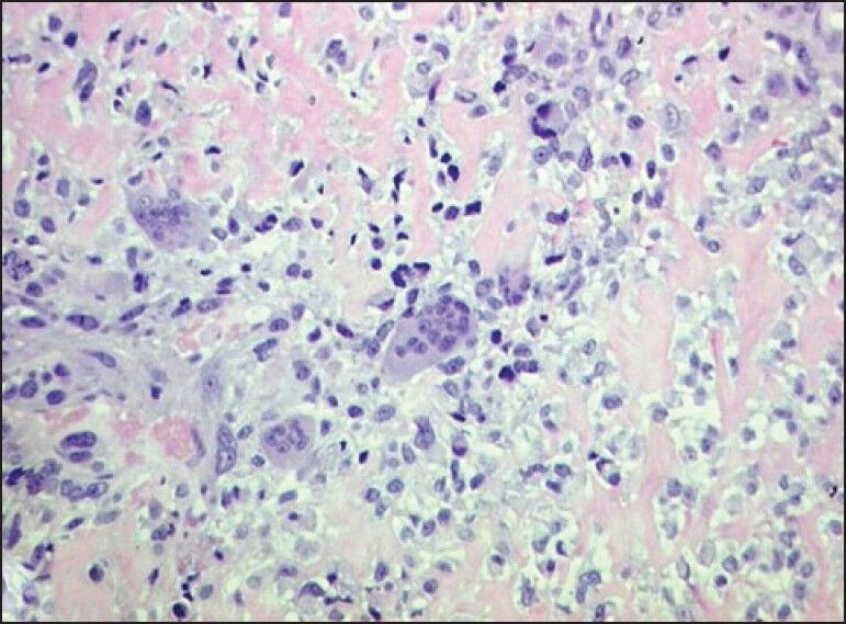
Photomicrograph of pathology section showing osteoclastic giant cells and pleomorphic malignant cells amidst osteoid (H and E, × 200)
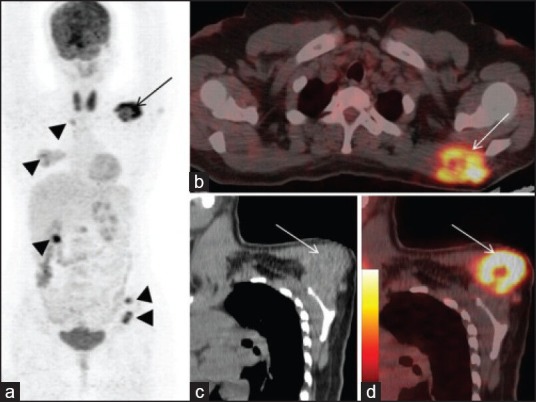
(a) Maximum intensity projection image showing tracer uptake in left supraclavicular region (2a-arrow), in the region of mid thorax, mid abdomen, right lung, right kidney, left pelvis (all arrow heads) (b) axial fused PET/CT images show FDG avid mass arising from left trapezius muscle (arrow) (c) oblique coronal CT and fused PET/CT (d) images show origin of the mass from left trapezius, with no involvement of left scapula
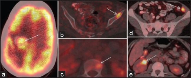
Axial fused PET/CT images show FDG avid metastatic lesions in brain (a – arrow), left iliac marrow (b – arrow), D12 vertebral body (c – arrow), left internal oblique muscle (d – arrow) and right renal cortex (e – arrow)
Similar articles
-
18F-FDG PET/CT in Isolated Primary Extraskeletal Osteosarcoma.Clin Nucl Med. 2018 Dec;43(12):e463-e464. doi: 10.1097/RLU.0000000000002293. Clin Nucl Med. 2018. PMID: 30273208
-
18F-FDG PET/CT in a rare malignant extraskeletal osteosarcoma.Clin Nucl Med. 2013 Sep;38(9):e367-9. doi: 10.1097/RLU.0b013e3182868ace. Clin Nucl Med. 2013. PMID: 23856822
-
(18)F-fluoro-deoxyglucose positron emission tomography-computed tomography in initial assessment and diagnosis of right atrial angiosarcoma with widespread visceral metastases: A rare case report and review of the literature.Indian J Nucl Med. 2015 Jan-Mar;30(1):51-4. doi: 10.4103/0972-3919.147541. Indian J Nucl Med. 2015. PMID: 25589807 Free PMC article.
-
Subcutaneous extraskeletal osteosarcoma of the forearm: a case report and review of the literature.Skeletal Radiol. 2016 Sep;45(9):1307-11. doi: 10.1007/s00256-016-2426-3. Epub 2016 Jun 30. Skeletal Radiol. 2016. PMID: 27357312 Review.
-
Primary extraskeletal osteosarcoma of sigmoid mesocolon: a case report and a review of the literature.World J Surg Oncol. 2021 Sep 3;19(1):267. doi: 10.1186/s12957-021-02337-9. World J Surg Oncol. 2021. PMID: 34479594 Free PMC article. Review.
Cited by
-
[Superficial extraskeletal osteosarcoma. Case report].Rev Med Inst Mex Seguro Soc. 2024 Mar 4;62(2):1-6. doi: 10.5281/zenodo.10712277. Rev Med Inst Mex Seguro Soc. 2024. PMID: 39514394 Free PMC article. Spanish.
-
Extraskeletal Osteosarcoma- A Case Report.J Clin Diagn Res. 2016 Jan;10(1):ED03-4. doi: 10.7860/JCDR/2016/16183.7065. Epub 2016 Jan 1. J Clin Diagn Res. 2016. PMID: 26894075 Free PMC article.
References
-
- Allan CJ, Soule EH. Osteogenic sarcoma of the somatic soft tissues. Clinicopathologic study of 26 cases and review of literature. Cancer. 1971;27:1121–33. - PubMed
-
- Eftekhari F. Imaging assessment of osteosarcoma in childhood and adolescence: Diagnosis, staging, and evaluating response to chemotherapy. Cancer Treat Res. 2009;152:33–62. - PubMed
-
- Bane BL, Evans HL, Ro JY, Carrasco CH, Grignon DJ, Benjamin RS, et al. Extraskeletal osteosarcoma. A clinicopathologic review of 26 cases. Cancer. 1990;65:2762–70. - PubMed
-
- Lee JS, Fetsch JF, Wasdhal DA, Lee BP, Pritchard DJ, Nascimento AG. A review of 40 patients with extraskeletal osteosarcoma. Cancer. 1995;76:2253–9. - PubMed
-
- Chung EB, Enzinger FM. Extraskeletal osteosarcoma. Cancer. 1987;60:1132–42. - PubMed
Publication types
LinkOut - more resources
Full Text Sources
Other Literature Sources
