. 2013 Dec;3(4):816-30.
doi: 10.1086/674753.

Nitrosation-dependent caveolin 1 phosphorylation, ubiquitination, and degradation and its association with idiopathic pulmonary arterial hypertension

Affiliations

Nitrosation-dependent caveolin 1 phosphorylation, ubiquitination, and degradation and its association with idiopathic pulmonary arterial hypertension

Farnaz R Bakhshi et al. Pulm Circ. 2013 Dec.

Abstract

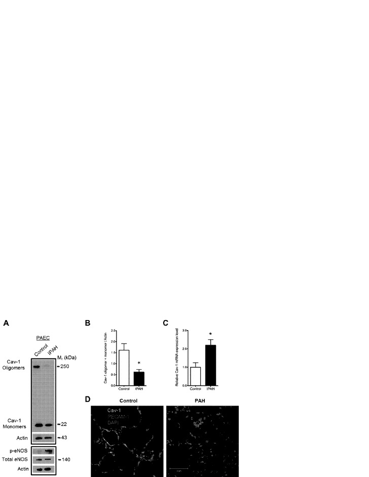
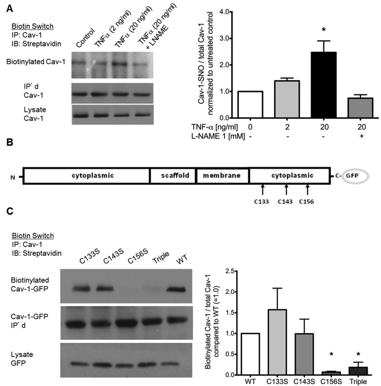
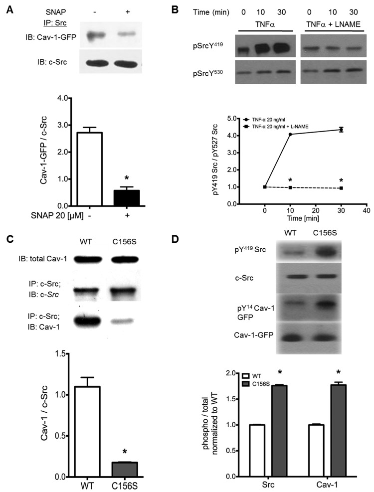
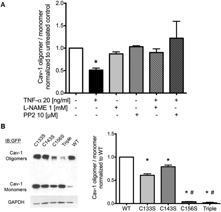
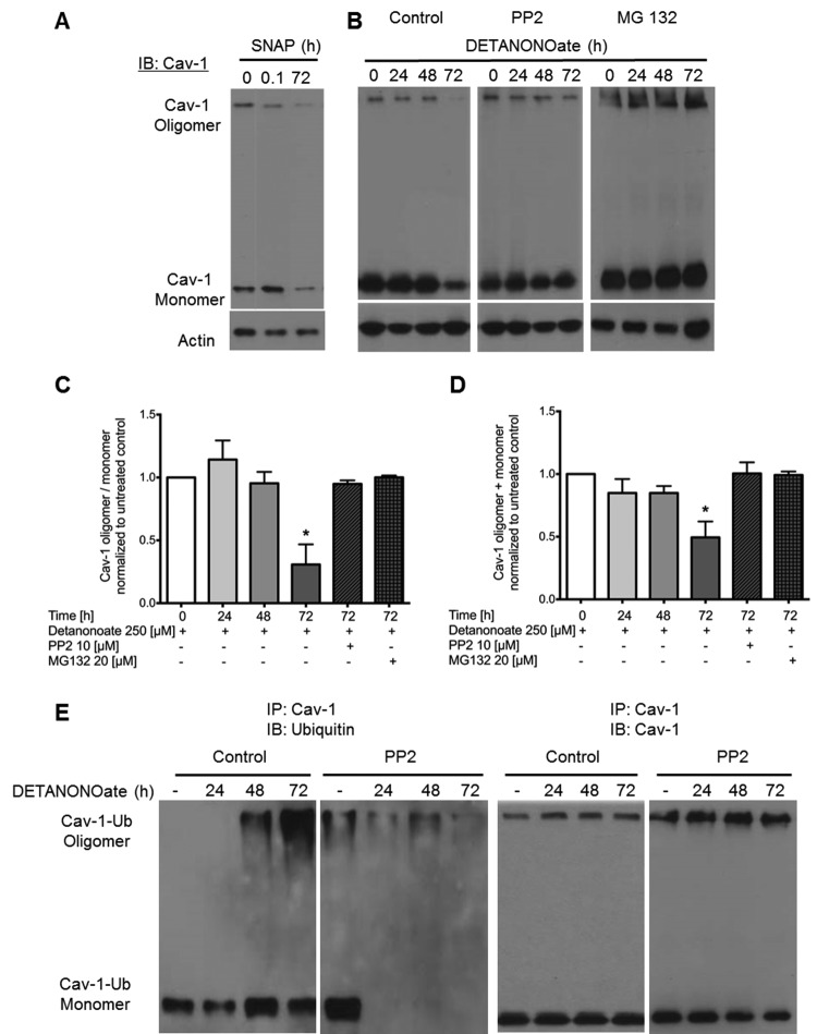
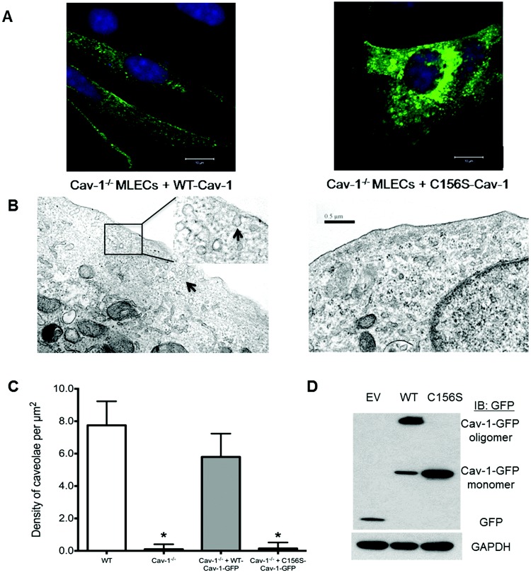
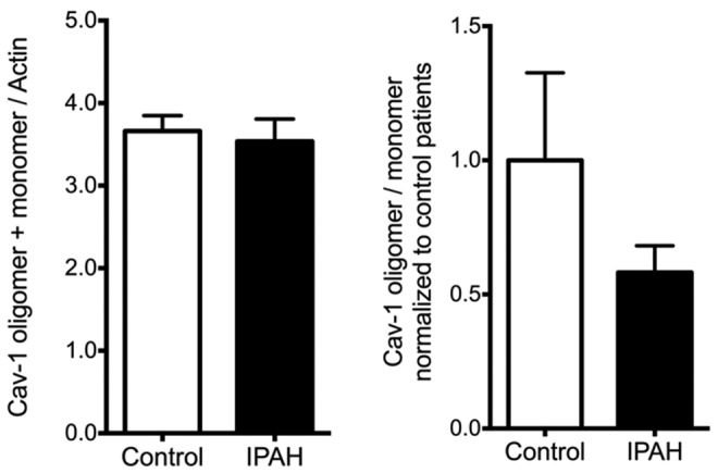
In the present study, we tested the hypothesis that chronic inflammation and oxidative/nitrosative stress induce caveolin 1 (Cav-1) degradation, providing an underlying mechanism of endothelial cell activation/dysfunction and pulmonary vascular remodeling in patients with idiopathic pulmonary arterial hypertension (IPAH). We observed reduced Cav-1 protein despite increased Cav-1 messenger RNA expression and also endothelial nitric oxide synthase (eNOS) hyperphosphorylation in human pulmonary artery endothelial cells (PAECs) from patients with IPAH. In control human lung endothelial cell cultures, tumor necrosis factor α-induced nitric oxide (NO) production and S-nitrosation (SNO) of Cav-1 Cys-156 were associated with Src displacement and activation, Cav-1 Tyr-14 phosphorylation, and destabilization of Cav-1 oligomers within 5 minutes that could be blocked by eNOS or Src inhibition. Prolonged stimulation (72 hours) with NO donor DETANONOate reduced oligomerized and total Cav-1 levels by 40%-80%, similar to that observed in IPAH patient-derived PAECs. NO donor stimulation of endothelial cells for >72 hours, which was associated with sustained Src activation and Cav-1 phosphorylation, ubiquitination, and degradation, was blocked by NOS inhibitor L-NAME, Src inhibitor PP2, and proteosomal inhibitor MG132. Thus, chronic inflammation, sustained eNOS and Src signaling, and Cav-1 degradation may be important causal factors in the development of IPAH by promoting PAEC dysfunction/activation via sustained oxidative/nitrosative stress.

Keywords: Src; endothelial dysfunction; endothelial nitric oxide synthase (eNOS); oxidative stress; pulmonary arterial hypertension (PAH).


Figures

Figure 1
Figure 1
Reduced caveolin 1 (Cav-1) oligomer and monomer protein level in pulmonary artery endothelial cells (PAECs) from patients with idiopathic pulmonary arterial hypertension (IPAH). A, Representative Western blot of PAEC lysates obtained from a healthy donor (left lane) and a patient with IPAH (right lane). The lower panel reveals an increase in phospho-Ser-1177 endothelial nitric oxide synthase (p-eNOS) relative to total eNOS expression in IPAH endothelial cells, indicative of eNOS hyperactivation/dysfunction. B, Summarized data. Asterisk indicates P < 0.05 versus control; n = 5. C, Quantitative polymerase chain reaction indicates increased Cav-1 messenger RNA (mRNA) in IPAH PAECs compared with control. Asterisk indicates P < 0.05; n = 3. D, Immunohistochemical staining and confocal microscopy of Cav-1 (green) and platelet endothelial cell adhesion molecule 1 (PECAM-1; red) in formalin-fixed, paraffin-embedded control and PAH lung sections. Note the significant colocalization of Cav-1 and PECAM-1 in control lung pulmonary arteriole and reduced Cav-1 staining in the endothelial cells of the remodeled pulmonary arteriole of the PAH lung section. Staining is representative of 6 PAH and control lung samples. Mr: relative molecular mass; DAPI: 4′,6-diamidino-2-phenylindole dihydrochloride.
Figure 2
Figure 2
Tumor necrosis factor α (TNF-α) induces caveolin 1 (Cav-1) S-nitrosation on Cys-156. A, Biotin switch assay indicates TNF-α-dependent, L-NAME-inhibitable nitrosation of Cav-1. TNF-α (2–20 ng/mL) was added to human lung microvascular endothelial cells (HLMVECs) 5 minutes before lysis. L-NAME was added 30 minutes prior to TNF-α. B, Biotin switch assay performed on human embryonic kidney cells overexpressing endothelial nitric oxide synthase that were expressing wild type (WT)–Cav-1, C133S-Cav-1, C143S-Cav-1, and C156S-Cav-1 mutants in pEGFP vector. C133S-, C143S-, C156S, and triple (C133S/C143S/C156S)–Cav-1 mutants in pEGFP vectors were made. C, Nitrosation was promoted by adding S-nitroso-N-acetyl-D,L-penicillamine (SNAP; 20 μM) at room temperature for 5 minutes. After lysis and the biotin switch, samples were immunoprecipitated with Cav-1 and blotted for streptavidin. Right panels show quantified densitometry of Western blots using ImageJ software. Bars show mean ± standard deviation. Asterisks indicate P < 0.05 versus control (A) or WT (C); n = 3. GFP: green fluorescent protein.
Figure 3
Figure 3
Nitric oxide (NO) donor and caveolin 1 (Cav-1)–C156S mutant decrease Cav-1/Src interaction and increase Src activity. A, Human embryonic kidney (HEK) cells were transfected with wild type (WT)–Cav-1–green fluorescent protein (GFP) and stimulated with either NO donor or S-nitroso-N-acetyl-D,L-penicillamine (SNAP; 20 μM, 5 minutes) or left untreated. Cells were lysed, Src was immunoprecipitated, and bound Cav-1 was quantified by Western blot. Asterisks indicate P < 0.05 versus control; n = 3. B, Analysis of active/inactive Src as indicated. pTyr419/pTyr530 Western blots from tumor necrosis factor α (TNF-α)–stimulated human lung microvascular endothelial cells with or without NO synthase (NOS) inhibitor pretreatment indicates that L-NAME blocks TNF-α-induced Src activation. Asterisks indicate P < 0.05 versus TNF-α alone; n = 3. C, HEK cells overexpressing endothelial NOS (eNOS; HEK-eNOS cells) were transfected with empty vector (EV), WT-Cav-1, and C156S-Cav-1 mutant in pEGFP vector. Samples were blotted for total Cav-1 (upper row) or immunoprecipitated with anti-Src monoclonal antibody and blotted for Cav-1 and Src (lower rows). D, HEK-eNOS cells were transfected with WT-Cav-1 and C156S-Cav-1 mutant and blotted for Src-pTyr418, total Src, Cav-1-pTyr14, and total Cav-1. Asterisks indicate P < 0.05 versus WT-Cav-1; n = 3.
Figure 4
Figure 4
Caveolin 1 (Cav-1) oligomerization is reduced by tumor necrosis factor α (TNF-α) in a nitric oxide (NO)– and Src-dependent manner. A, Human lung microvascular endothelial cells were pretreated with either nitric oxide synthase inhibitor L-NAME or Src inhibitor PP2 30 minutes prior to being challenged with TNF-α for 5 minutes. Cav-1 oligomer/monomer distribution was analyzed by Western blot of nonboiled samples. Quantification of densitometry using ImageJ (mean ± SD; asterisk indicates P < 0.05 versus untreated; n = 3). B, Western blot analysis of Cav-1 expressed in human embryonic kidney cells (EV = empty green fluorescent protein [GFP] vector, wild type [WT]–, C133S-, C143S-, C156S-, and triple [C133S/C143S/C156S]–Cav-1-GFP mutants) showed that Cys-156 is required to maintain oligomer stability. Bar graphs show quantified densitometry of Cav-1 oligomer/monomer. Asterisks indicate P < 0.05 versus WT-Cav-1; pound signs indicate P < 0.05 versus C133S- and C143S-Cav-1; n = 3.
Figure 5
Figure 5
Nitric oxide (NO) donors induce Src-dependent caveolin 1 (Cav-1) oligomer destabilization and Cav-1 degradation. A, Human lung microvascular endothelial cells (HLMVECs) treated with S-nitroso-N-acetyl-D,L-penicillamine (SNAP; 10 μM) showed reduced oligomerization at 5 minutes (0.08 hours) and a decrease in total Cav-1 expression at 72 hours. B, HLMVECs either untreated or pretreated with the Src inhibitor PP2 (10 μM) or the proteosome inhibitor MG132 (20 μm) were challenged every 24 hours with NO donor DETANONOate (250 μM) for 24, 48, or 72 hours. Nonboiled samples were separated by sodium dodecyl sulfate polyacrylamide gel electrophoresis and blotted for Cav-1 and actin. C, D, Quantified densitometry of Western blots reveal a significant decrease in the Cav-1 oligomer/monomer ratio (C) and a significant decrease in total Cav-1 (monomer + oligomer; D) after 72 hours of DETANONOate, which is blocked by PP2 and MG132. Asterisks indicate P < 0.05 versus untreated by analysis of variance; n = 3. E, Ubiquitination of immunoprecipitated Cav-1 from HLMVEC lysates after challenge with NO donor DETANONOate (250 μM) in the presence or absence of PP2 (10 μM; left panels) and reprobe of membrane with anti-Cav-1 antibody (right panels). Immunoblots show that ubiquitination (left panel) and degradation (right panel) of Cav-1 (oligomers and monomers) following treatment with DETANONOate is blocked by PP2 (representative of 3 experiments).
Figure 6
Figure 6
Confocal and transmission electron microscopy reveals that the C156S caveolin 1 (Cav-1) mutant fails to rescue caveolae formation in Cav-1−/− mouse lung endothelial cells (MLECs). A, Cav-1−/− MLECs transfected with wild type (WT)–Cav-1–green fluorescent protein (GFP) complementary DNA (cDNA) restored membrane-associated Cav-1 localization, whereas the C156S-Cav-1-GFP mutant accumulated in perinuclear intracellular compartments. B, Representative transmission electron microscopy micrographs show rescue of caveolae in Cav-1−/− endothelial cells following transduction of WT-Cav-1, whereas no caveolae were observed in Cav1−/− MLECs transduced with C156S-Cav-1 cDNA. C, Quantitative analysis of electron micrographs indicate near-complete rescue of caveolae formation by WT-Cav-1 compared with WT control and Cav-1−/− mouse lung endothelial cells and lack of caveolae formation in cells transduced with C156S-Cav-1. Asterisks indicate P < 0.05 versus WT control MLECs by analysis of variance; n = 10 sections per group. D, Western blot analysis of WT-Cav-1-GFP, C156S-Cav-1-GFP, and empty GFP vector (EV) expression in human embryonic kidney cells. Note the absence of stable high-molecular-weight oligomers in C156S-Cav-1-transfected cells.
Figure S1
Figure S1
Human pulmonary microvascular endothelial cell (HPMVEC) lysates showed no change in total caveolin 1 (Cav-1) protein expression, but there was a trend toward reduced oligomerization in pulmonary arterial hypertension (PAH) samples. IPAH: idiopathic PAH.
Figure S2
Figure S2
Three unique peptides from caveolin 1 (Cav-1) were identified by liquid chromatography tandem mass spectrometry (MS/MS) analysis of the 50-kDa Cav-1–green fluorescent protein (GFP) agarose gel band. The MS/MS spectrum of an ion at mass 860.67 (4+) was matched by Mascot software to Cav-1 tryptic peptide 66–86 IDFEDVIAEPEGTHSFDGIWK containing one ubiquitin group. The measured mass for this peptide was 3,438.63 Da, which is −1.8 ppm different from the theoretical value. The site of ubiquitination modification is indicated by the fragment ion at mz 1,324.1, which corresponds to the y(22+2H) ion with a modified lysine. The lysine is residue 86 in the Cav-1 sequence.

References

    1. Maniatis N, Chernaya O, Shinin V, Minshall R. Caveolins and lung function. Adv Exp Med Biol 2012;729:157–179. - PMC - PubMed
    1. Stan R-V. Structure and function of endothelial caveolae. Microsc Res Tech 2002;57:350–364. - PubMed
    1. Minshall RD, Sessa WC, Stan RV, Anderson RGW, Malik AB. Caveolin regulation of endothelial function. Am J Physiol Lung Cell Mol Physiol 2003;285:L1179–L1183. - PubMed
    1. Stan RV. Structure of caveolae. Biochim Biophys Acta 2005;1746:334–348. - PubMed
    1. Rothberg KG, Heuser JE, Donzell WC, Ying Y-S, Glenney JR, Anderson RGW. Caveolin, a protein component of caveolae membrane coats. Cell 1992;68:673–682. - PubMed