Piglet model of chronic pulmonary hypertension
- PMID: 25006407
- PMCID: PMC4070840
- DOI: 10.1086/674757
Piglet model of chronic pulmonary hypertension
Abstract
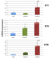
None of the animal models have been able to reproduce all aspects of CTEPH because of the rapid resolution of the thrombi in the pulmonary vasculature. The aim of this study was to develop an easily reproducible large-animal model of chronic pulmonary hypertension (PH) related to the development of a postobstructive and overflow vasculopathy. Chronic PH was induced in 5 piglets by ligation of the left pulmonary artery (PA) through a midline sternotomy followed by weekly transcatheter embolization of the right lower-lobe arteries. Sham-operated piglets (n = 5) served as controls. Hemodynamics, RV function, lung morphometry, and endothelin-1 (ET-1) pathway gene expression (ET-1 and its receptors ETA and ETB) were assessed after 5 weeks in the obstructed (left lung and right lower lobe) and unobstructed (right upper lobe) territories. All animals developed chronic PH within 5 weeks. Compared to controls, chronic-PH animals had higher mean PA pressure (28.5 ± 1.7 vs. 11.6 ± 1.8 mmHg, P = 0.0001) and total pulmonary resistance (784 ± 160 vs. 378 ± 51 dyn s(-1) cm(-5), P = 0.05). Echocardiography showed RV enlargement, RV wall thickening (56 ± 5 vs. 30 ± 4 mm, P = 0.0003), decreased tricuspid annular plane systolic excursion (11.3 ± 0.9 vs. 14.4 ± 0.4 mm, P = 0.01), and paradoxical septal motion. In obstructed territories, morphometry demonstrated increases in the number of bronchial arteries per bronchus (8.7 ± 0.9 vs. 2 ± 0.17, P < 0.0001) and in distal PA media thickness (60% ± 2.8% vs. 29% ± 0.9%, P < 0.0001), consistent with postobstructive vasculopathy. Distal PA media thickness was increased in unobstructed territories (70% ± 2.4% vs. 29% ± 0.9%, P < 0.0001). ET-1 was overexpressed in unobstructed territories, compared to controls and obstructed territories. In conclusion, the large-animal model described here is reproducible and led to the development of PH in a relatively short time frame.
Keywords: animal model; chronic thromboembolic pulmonary hypertension; pulmonary hypertension.
Figures


References
-
- Simonneau G, Robbins IM, Beghetti M, Channick RN, Delcroix M, Denton CP, Elliott CG, et al. Updated clinical classification of pulmonary hypertension. J Am Coll Cardiol 2009;54(1_suppl):S43–S54. - PubMed
-
- Dartevelle P, Fadel E, Mussot S, Chapelier A, Hervé P, de Perrot M, Cerrina J, et al. Chronic thromboembolic pulmonary hypertension. Eur Respir J 2004;23(4):637–648. - PubMed
-
- Galiè N, Kim N. Pulmonary microvascular disease in chronic thromboembolic pulmonary hypertension. Proc Am Thorac Soc 2006;3:571–576. - PubMed
-
- Hoeper MM, Mayer E, Simonneau G, Rubin LJ. Chronic thromboembolic pulmonary hypertension. Circulation 2006;113(16):2011–2020. - PubMed
-
- Moser KM, Bloor CM. Pulmonary vascular lesions occurring in patients with chronic major vessel thromboembolic pulmonary hypertension. Chest 1993;103(3):685–692. - PubMed
LinkOut - more resources
Full Text Sources
Other Literature Sources

