

A unique pulmonary microvascular endothelial cell niche revealed by Weibel-Palade bodies and Griffonia simplicifolia
- PMID: 25006426
- PMCID: PMC4070765
- DOI: 10.1086/674879
A unique pulmonary microvascular endothelial cell niche revealed by Weibel-Palade bodies and Griffonia simplicifolia
Abstract
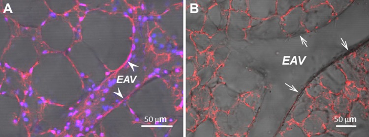
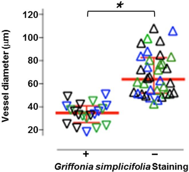
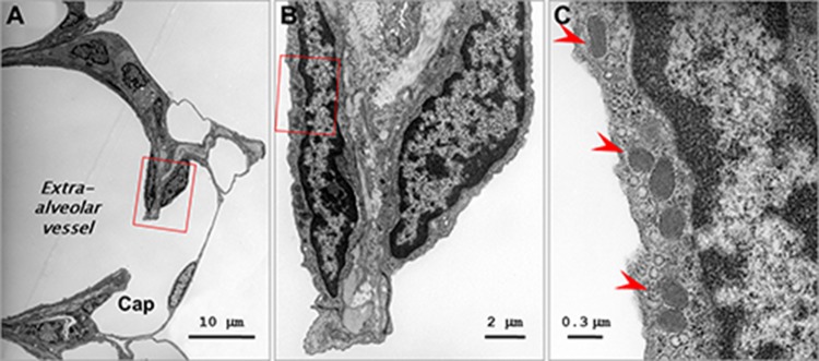
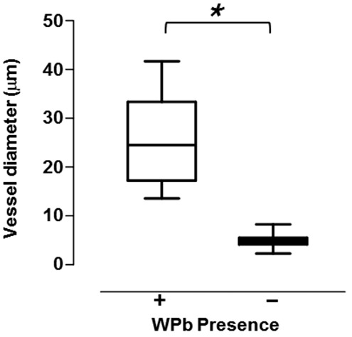
Pulmonary endothelium displays considerable heterogeneity along the vascular axis, from arteries to capillaries to veins. Griffonia simplicifolia is a lectin that recognizes pulmonary microvascular endothelium with preference over extra-alveolar endothelium in both arteries and veins, yet the precise vascular location where this phenotypic shift occurs is poorly resolved. We gelatin-filled the circulation and agarose-loaded the airways and then labeled the lung with Griffonia lectin to enable visualization of the endothelial transition zone. Endothelium in vessels with internal diameters less than 38 μm were uniformly Griffonia positive, whereas vessels with internal diameters greater than 60 μm were always Griffonia negative. Two populations of endothelium were identified in vessels ranging from 38 to 60 μm in diameter, including some that were positive and others that were negative for binding to G. simplicifolia. To better resolve this endothelial transition zone, we performed morphology studies to measure the distribution of Weibel-Palade bodies (WPbs), since WPbs are present in conduit vessel endothelium and absent in capillary endothelium. WPbs were found in endothelium with vascular dimensions as small as 18 μm in diameter but were not found in capillaries. Thus, we identify with precision that the endothelial phenotype transition from a cell that does not interact with Griffonia lectin to one that does occurs in blood vessels with internal diameters of approximately 38 μm, and we reveal an unappreciated vascular zone, between 18 and 38 μm in diameter, where endothelium both is Griffonia positive and possesses WPbs.
Keywords: P-selectin; heterogeneity; lectin; vascular; von Willebrand factor.
Figures
References
-
- Laubichler MD, Aird WC, Maienschein J. The endothelium in history. In: Aird WC, ed. Endothelial biomedicine. Cambridge: Cambridge University Press, 2007:5–19.
-
- Aird WC. Phenotypic heterogeneity of the endothelium. II. Representative vascular beds. Circ Res 2007;100:174–190. - PubMed
-
- Aird WC. Phenotypic heterogeneity of the endothelium. I. Structure, function, and mechanisms. Circ Res 2007;100:158–173. - PubMed
-
- Stevens T. Microheterogeneity of the lung endothelium. In: Aird WC, ed. Endothelial biomedicine. Cambridge: Cambridge University Press; 2007:1161–1170.
-
- Gebb S, Stevens T. On lung endothelial cell heterogeneity. Microvasc Res 2004;68:1–12. - PubMed
Grants and funding
LinkOut - more resources
Full Text Sources
Other Literature Sources
