. 2014 Mar;4(1):116-27.
doi: 10.1086/675641.

The serine-threonine phosphatase calcineurin is a regulator of endothelial store-operated calcium entry

Affiliations

The serine-threonine phosphatase calcineurin is a regulator of endothelial store-operated calcium entry

Audrey A Vasauskas et al. Pulm Circ. 2014 Mar.

Abstract

Disruption of the endothelium leads to increased permeability, allowing extravasation of macromolecules and other solutes from blood vessels. Calcium entry through a calcium-selective, store-operated calcium (SOC) channel, I soc, contributes to barrier disruption. An understanding of the mechanisms surrounding the regulation of I soc is far from complete. We show that the calcium/calmodulin-activated phosphatase calcineurin (CN) plays a role in regulation of SOC entry, possibly through the dephosphorylation of stromal interaction molecule 1 (STIM1). Phosphorylation has been implicated as a regulatory mechanism of activity for a number of canonical transient receptor potential (TRPC) and SOC channels, including I soc. Our results show that STIM1 phosphorylation increases in pulmonary artery endothelial cells (PAECs) upon activation of SOC entry. However, the phosphatases involved in STIM1 dephosphorylation are unknown. We found that a CN inhibitor (calcineurin inhibitory peptide [CIP]) increases the phosphorylation pattern of STIM1. Using a fura 2-acetoxymethyl ester approach to measure cytosolic calcium in PAECs, we found that CIP decreases SOC entry following thapsigargin treatment in PAECs. Luciferase assays indicate that thapsigargin induces activation of CN activity and confirm inhibition of CN activity by CIP in PAECs. Also, I soc is significantly attenuated in whole-cell patch-clamp studies of PAECs treated with CIP. Finally, PAECs pretreated with CIP exhibit decreased interendothelial cell gap formation in response to thapsigargin-induced SOC entry, as compared to control cells. Taken together, our data show that CN contributes to the phosphorylation status of STIM1, which is important in regulation of endothelial SOC entry and I soc activity.

Keywords: Isoc; STIM1; TRPC; phosphatase; phosphorylation; stromal interaction molecule 1; transient receptor potential.


Figures
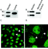
Figure 1
Figure 1
Localization of calcineurin (CN) in pulmonary artery endothelial cells (PAECs). A, Western blot analysis of CN in whole-cells lysates of PAECs. Whole-cell lysates of PAECs were run on 4%–12% bis-tris gels, transferred onto nitrocellulose, and subjected to Western blot analysis with antibody to pan-CN A. A ∼57-kDa band corresponding to CN A was observed. B, Immunoblot showing CN in supernatant detergent-extracted membrane fraction using octylglucoside/KI. CN A was found in the membrane fraction of PAECs. C, Immunocytochemical staining for CN in PAECs. Fluorescent images were obtained with a Leica TCS SP2 confocal laser scanning microscope fitted with a 63× water immersion objective. Arrows highlight membrane localization of positive CN staining. D, Enlarged detail of cell-cell border staining in C.
Figure 2
Figure 2
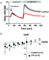
Calcineurin inhibitory peptide (CIP) attenuates store-operated calcium (SOC) entry and decreases ISOC in pulmonary artery endothelial cells (PAECs). A, PAECs were loaded with Fura 2/AM (fura 2-acetoxymethyl ester) and pretreated with 50 μM CIP or vehicle alone for 1 hour, and changes in intracellular [Ca2+] were measured. SOC entry was revealed by thapsigargin treatment in low-Ca2+ buffer. Upon 2 mM Ca2+ add-back, CIP-treated PAECs exhibited a decreased SOC (red tracing) compared to controls (black tracing). Each tracing represents mean ± SEM of at least 4 experiments with 2 regions of interest of 10–20 cells for each. B, Single cells were pretreated with CIP for 1–2 hours and examined in a whole-cell patch-clamp configuration with CIP added to the patch pipette. The ISOC channel was activated by thapsigargin in the patch pipette. Cells were held at 0 mV and stepped from −100 to +100 mV for 200 milliseconds. CIP treatment attenuated ISOC at all negative testing potentials, as compared to control cells. Asterisk indicates significant differences from control cells.
Figure 3
Figure 3
Calcineurin inhibitory peptide (CIP) reduces thapsigargin (TG)-induced calcineurin (CN) activity in pulmonary artery endothelial cells (PAECs). PAECs were transiently transfected with a nuclear factor of activated T cells (NFAT)–responsive luciferase reporter plasmid, an indicator of CN phosphatase activity. CN activity is shown as relative light units (RLU) of luciferase normalized to total protein. CN activity increased upon TG treatment, and this increase was attenuated in CIP-treated cells. Each bar represents the mean + SEM of at least 3 independent experiments. Asterisk indicates significant differences from TG-treated cells.
Figure 4
Figure 4
Stromal interaction molecule 1 (STIM1) is phosphorylated in pulmonary artery endothelial cell (PAECs). A, Western blot analysis of phosphoserine-immunoreactive proteins in whole-cell lysates of PAECs. Whole-cell lysates of PAECs treated with and without thapsigargin (+TG and −TG, respectively) for 1, 3, and 5 minutes were run on 4%–12% bis-tris gels, transferred onto nitrocellulose, and subjected to Western blot analysis with antibody to phosphoserine residues. An ∼85-kDa band was observed that was increased upon TG treatment. Actin is shown as loading control. Below, independent immunoblot of whole-cell lysates, showing STIM1 immunoreactivity at ∼85 kDa by use of a polyclonal antibody to STIM1. B, STIM1 was immunoprecipitated from PAEC whole-cell lysates, subjected to Western blot analysis, and probed for phosphoserine immunoreactivity. A band corresponding to phospho-STIM1 at ∼85 kDa was observed. E1, elution, STIM1 immunoprecipitate. C, Western blot analysis of phosphoserine-immunoreactive proteins in whole-cell lysates of PAECs treated with or without thrombin (+Th and −Th, respectively) for 1, 3, 5, 7, and 10 minutes and run on 4%–12% bis-tris gels, transferred onto nitrocellulose, and subjected to Western blot analysis with antibody to phosphoserine residues. Actin is shown as loading control.
Figure 5
Figure 5
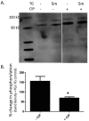
Calcineurin inhibitory peptide (CIP) increases phosphorylation of stromal interaction molecule 1 (STIM1) in pulmonary artery endothelial cells (PAECs). A, Western blot analysis of CIP-treated cells. Supernatant octylglucoside/KI-extracted membrane fractions of cells treated with or without 5-minute thapsigargin (TG) treatment and with or without CIP and subjected to Western blot analysis with antiphosphoserine antibody. A phosphospecific band at ∼85 kDa was observed that increased in intensity with TG and/or CIP treatment. B, Western blot densitometry results from A. ImageJ software was used to measure the densitometry of bands. Data are represented as percent change in phosphorylation, based on band density of TG-treated cells or untreated cells. Each bar represents the mean ± SEM of 2 independent membrane preparations. Asterisk indicates significant difference from non-CIP-treated controls.
Figure 6
Figure 6
Stromal interaction molecule 1 (STIM1) knockdown attenuates store-operated calcium (SOC) in pulmonary artery endothelial cells (PAECs). PAECs were transfected with 20 nM siRNA (small interfering RNA) to knockdown STIM1 using HiPerFect transfection reagent. Control cells received 20 nM scrambled (Scram) siRNA. A, Representative Western blots showing STIM1 levels in PAECs with and without siRNA treatment. Actin is shown as loading control. B, Densitometry analysis of siRNA-treated PAECs. An asterisk indicates significant differences from non-siRNA-treated control cells. C, Thapsigargin-induced SOC entry was decreased in PAECs treated with siRNA- and CIP-treated cells. PAECs received 20 nM of siRNA to knockdown STIM1 for >72 hours (or scrambled control). Cells were loaded with Fura 2/AM (fura 2-acetoxymethyl ester) and pretreated with 50 μM calcineurin inhibitory peptide (CIP) or vehicle alone for 1 hour, and changes in intracellular [Ca2+] were measured. SOC entry was initiated by thapsigargin treatment in low-Ca2+ buffer. Upon 2 mM Ca2+ add-back, cells treated with siRNA to STIM1 exhibited decreased SOC (red tracing) compared to controls (black tracing). CIP treatment had no effect on this decrease in SOC entry in STIM1 siRNA-treated cells (green tracing). Each tracing represents the average of at least 3 experiments (mean ± SEM) with 2 regions of interest of 10–20 cells for each. D, Graphical representation of calcium tracings in C at peak SOC entry (600 seconds).
Figure 7
Figure 7
Calcineurin inhibitory peptide (CIP) reduces thapsigargin-induced intercellular gap formation in pulmonary artery endothelial cells (PAECs). A, PAECs grown on glass coverslips exhibit confluent monolayer at time 0. B, Thapsigargin treatment induces intercellular gap formation, as denoted by arrows. C, PAECs exhibit confluent monolayer after 1-hour pretreatment with 50 μM CIP. D, CIP-pretreated cells do not form intercellular gaps after thapsigargin treatment.

Similar articles

Cited by

References

    1. Parekh AB, Putney JW Jr. Store-operated calcium channels. Physiol Rev 2005;85:757–810. - PubMed
    1. Cioffi DL, Wu S, Stevens T. Transient receptor potential cation channels and store-operated Ca2+ channels. In: Yuan JX-J, ed. Ion channels in pulmonary vasculature. Lung Biology in Health and Disease, no. 197. Boca Raton, FL: Taylor & Francis, 2005:125–146.
    1. Wu S, Cioffi EA, Alvarez D, Sayner SL, Chen H, Cioffi DL, King J, et al. Essential role of a Ca2+-selective, store-operated current (ISOC) in endothelial cell permeability: determinants of the vascular leak site. Circ Res 2005;96:856–863. - PubMed
    1. Cioffi DL, Lowe K, Alvarez DF, Barry C, Stevens T. TRPing on the lung endothelium: calcium channels that regulate barrier function. Antioxid Redox Signal 2009;11:765–776. - PMC - PubMed
    1. Brough GH, Wu S, Cioffi D, Moore TM, Li M, Dean N, Stevens T. Contribution of endogenously expressed Trp1 to a Ca2+-selective, store-operated Ca2+ entry pathway. FASEB J 2001;15:1727–1738. - PubMed

LinkOut - more resources