

Could (18) F-DPA-714 PET imaging be interesting to use in the early post-stroke period?
- PMID: 25006546
- PMCID: PMC4077629
- DOI: 10.1186/s13550-014-0028-4
Could (18) F-DPA-714 PET imaging be interesting to use in the early post-stroke period?
Abstract
Background: Cerebral stroke is a severe and frequent condition that requires rapid and reliable diagnosis. If administered shortly after the first symptoms manifest themselves, IV thrombolysis has been shown to increase the functional prognosis by restoring brain reperfusion. However, a better understanding of the pathophysiology of stroke should help to identify potential new therapeutic targets. Stroke is known to induce an inflammatory brain reaction that involves overexpression of the 18-kDa translocator protein (TSPO) in glial cells and infiltrated leukocytes, which can be visualised by positron emission tomography (PET). We aimed to evaluate post-stroke neuroinflammation using the PET TSPO radioligand (18) F-DPA-714.
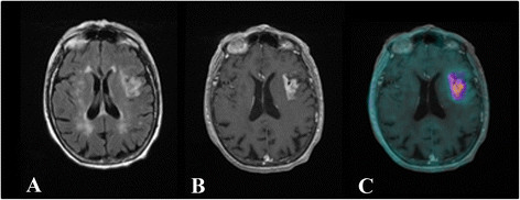
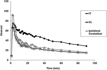
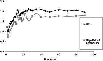
Methods: Nine patients underwent (18) F-DPA-714 PET and magnetic resonance imaging (MRI) between 8 and 18 days after the ictus. Co-registration of MRI and PET images was used to define three volumes of interest (VOIs): core infarction, contralateral region, and cerebellum ipsilateral to the stroke lesion. Time activity curves were obtained from each VOI, and ratios of mean and maximum activities between the VOIs were calculated.
Results: We observed an increased uptake of (18) F-DPA-714 co-localised with the infarct tissue and extension beyond the region corresponding to the damage in the blood brain barrier. No correlation was identified between (18) F-DPA-714 uptake and infarct volume. (18) F-DPA-714 uptake in ischemic lesion (mainly associated with TSPO expression in the infarct area and in the surrounding neighbourhood) slowly decreased from 10 min pi to the end of the PET acquisition, remaining higher than that in both contralateral region and ipsilateral cerebellum.
Conclusion: Our results show that (18) F-DPA-714 uptake after acute ischemia is mainly associated with TSPO expression in the infarct area and in the surrounding neighbourhood. We also demonstrated that the kinetics of (18) F-DPA-714 differs in injured tissue compared to normal tissue. Therefore, (18) F-DPA-714 may be useful in assessing the extent of neuroinflammation associated with acute stroke and could also help to predict clinical outcomes and functional recovery, as well as to assess therapeutic strategies, such as the use of neuroprotective/anti-inflammatory drugs.
Keywords: 18 F-DPA-714; Neuroinflammation; PET; Stroke.
Figures
Similar articles
-
11C-DPA-713 Versus 18F-GE-180: A Preclinical Comparison of Translocator Protein 18 kDa PET Tracers to Visualize Acute and Chronic Neuroinflammation in a Mouse Model of Ischemic Stroke.J Nucl Med. 2019 Jan;60(1):122-128. doi: 10.2967/jnumed.118.209155. Epub 2018 Jul 5. J Nucl Med. 2019. PMID: 29976695 Free PMC article.
-
Characterization of the inflammatory post-ischemic tissue by full volumetric analysis of a multimodal imaging dataset.Neuroimage. 2020 Nov 15;222:117217. doi: 10.1016/j.neuroimage.2020.117217. Epub 2020 Jul 31. Neuroimage. 2020. PMID: 32745676
-
Impact of hydroxytyrosol on stroke: tracking therapy response on neuroinflammation and cerebrovascular parameters using PET-MR imaging and on functional outcomes.Theranostics. 2021 Feb 15;11(9):4030-4049. doi: 10.7150/thno.48110. eCollection 2021. Theranostics. 2021. PMID: 33754046 Free PMC article.
-
Positron emission tomography to image cerebral neuroinflammation in ischaemic stroke: a pilot study.Southampton (UK): NIHR Journals Library; 2020 Feb. Southampton (UK): NIHR Journals Library; 2020 Feb. PMID: 32023020 Free Books & Documents. Review.
-
A mini-review: Application of the radiopharmaceutical [18F]DPA-714 in neuroinflammation research in post-SARS-CoV-2 patients.J Neurol Sci. 2025 Jun 15;473:123518. doi: 10.1016/j.jns.2025.123518. Epub 2025 Apr 28. J Neurol Sci. 2025. PMID: 40315803 Review.
Cited by
-
In vivo assessment of neuroinflammation in progressive multiple sclerosis: a proof of concept study with [18F]DPA714 PET.J Neuroinflammation. 2018 Nov 13;15(1):314. doi: 10.1186/s12974-018-1352-9. J Neuroinflammation. 2018. PMID: 30424780 Free PMC article.
-
Detection of neuroinflammation before selective neuronal loss appearance after mild focal ischemia using [18F]DPA-714 imaging.EJNMMI Res. 2018 Jun 8;8(1):43. doi: 10.1186/s13550-018-0400-x. EJNMMI Res. 2018. PMID: 29884977 Free PMC article.
-
Immune interventions in stroke.Nat Rev Neurol. 2015 Sep;11(9):524-35. doi: 10.1038/nrneurol.2015.144. Epub 2015 Aug 25. Nat Rev Neurol. 2015. PMID: 26303850 Free PMC article. Review.
-
Evaluation of the novel TSPO radiotracer [18F] VUIIS1008 in a preclinical model of cerebral ischemia in rats.EJNMMI Res. 2017 Nov 25;7(1):93. doi: 10.1186/s13550-017-0343-7. EJNMMI Res. 2017. PMID: 29177913 Free PMC article.
-
PET imaging of neuroinflammation in neurological disorders.Lancet Neurol. 2020 Nov;19(11):940-950. doi: 10.1016/S1474-4422(20)30346-X. Lancet Neurol. 2020. PMID: 33098803 Free PMC article. Review.
References
LinkOut - more resources
Full Text Sources
Other Literature Sources
Research Materials
Miscellaneous
