. 2013 Dec 5:2013:598587.
doi: 10.1155/2013/598587. eCollection 2013.

Competitive HIF Prolyl Hydroxylase Inhibitors Show Protection against Oxidative Stress by a Mechanism Partially Dependent on Glycolysis

Affiliations

Competitive HIF Prolyl Hydroxylase Inhibitors Show Protection against Oxidative Stress by a Mechanism Partially Dependent on Glycolysis

Ann-Louise Bergström et al. ISRN Neurosci. .

Abstract

The hypoxia inducible factor 1 (HIF-1) is a central transcription factor involved in the cellular and molecular adaptation to hypoxia and low glucose supply. The level of HIF-1 is to a large degree regulated by the HIF prolyl hydroxylase enzymes (HPHs) belonging to the Fe(II) and 2-oxoglutarate-dependent dioxygenase superfamily. In the present study, we compared competitive and noncompetitive HPH-inhibitor compounds in two different cell types (SH-SY5Y and PC12). Although the competitive HPH-inhibitor compounds were found to be pharmacologically more potent than the non-competitive compounds at inhibiting HPH2 and HPH1, this was not translated into the cellular effects of the compounds, where the non-competitive inhibitors were actually more potent than the competitive in stabilizing and translocatingHIF1 α to the nucleus (quantified with Cellomics ArrayScan technology). This could be explained by the high cellular concentrations of the cofactor 2-oxoglutarate (2-OG) as the competitive inhibitors act by binding to the 2-OG site of the HPH enzymes. Both competitive and non-competitive HPH inhibitors protected the cells against 6-OHDA induced oxidative stress. In addition, the protective effect of a specific HPH inhibitor was partially preserved when the cells were serum starved and exposed to 2-deoxyglucose, an inhibitor of glycolysis, indicating that other processes than restoring energy supply could be important for the HIF-mediated cytoprotection.


Figures

Figure 1
Figure 1
Cellomics images showing the algorithm used for quantification of nuclear and cytoplasmic levels of HIF-1α. Raw images without the applied algorithm used to define cells are shown in the top panel and images showing the algorithm are shown in the lower panel. Circ is defined by the outline of the Hoechst-staining and thus represents the nuclear region. Ring is defined as a certain radius surrounding the Circ region and thus represents the cytoplasmic region of the cell. The Hoechst fluorescence and the HIF-1α fluorescence are recorded in two different channels.
Figure 2
Figure 2
FG41, DFO, CpdA, and JNJ induce HIF-1α upregulation and nuclear translocation in SH-SY5Y cells. (a) Immunocytochemical detection of HIF-1α in SH-SY5Y cells after 3 hours of treatment with FG41 or CpdA (both at 25 μM). The top panel shows HIF-1α immunoreactivity alone and the lower panel shows HIF-1α merged with Hoechst. Purple nuclei in the merged image indicate nuclear localization of HIF-1α. (b) Cellomics ArrayScan quantification of the total nuclear level of HIF-1α induced after 3 or 24 hours of FG41 or DFO treatment (both at 25 μM). The levels are represented as percentage of control (untreated). (c) Cellomics ArrayScan quantification of the ratio of nuclear to cytosolic located HIF-1α (percentage of untreated control) after FG41 or DFO treatment as in (b). (d) Nuclear HIF-1α after treatment with CpdA or JNJ (both 25 μM). (e) Nuclear/cytosolic ratio of HIF-1α after treatment with CpdA or JNJ as in (e). Asterisks indicate levels significantly different from control in a t-test.
Figure 3
Figure 3
HIF-1α stabilization and nuclear translocation in differentiated SHSH-5Y cells. The cells were treated with 10 or 25 μM FG41 for 3 hours, then fixed, and Hoechst-stained. The upper panel shows HIF-1α immunoreactivity alone and the lower panel shows the images merged with the Hoechst-staining.
Figure 4
Figure 4
Neuroprotective effect of HPH inhibitors. (a) ATP-levels in SH-SY5Y cells grown overnight in starvation media, pretreated for 3 hours with 25 μM of FG41 (a), DFO (b), CpdA (c), or JNJ (d) followed by 6-OHDA treatment for 3 hours. (e) Mitochondrial membrane potential in SH-SY5Y cells treated with Cpd A (10 or 25 μM) for 3 hours followed by 3 hours treatment with 50 μM 6-OHDA. Asterisks indicate levels significantly increased (t-test) as compared to control.
Figure 5
Figure 5
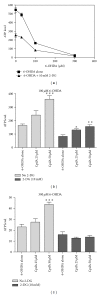
Cpd A is partially protective even when glycolysis is inhibited. (a) ATP-levels in SH-SY5Y cells grown overnight in starvation media overnight, then treated with 10 mM 2-deoxyglucose for 3 hours (or nothing for control) followed by 3 hours of treatment with 6-OHDA at the indicated concentrations. (b) and (c) ATP-levels in SH-SY5Y cells grown over night in starvation media, then pretreated for 3 hours with 10 mM 2-deoxyglucose (or nothing for control) and Cpd A (25 or 50 μM or nothing for control) followed by 3 hours treatment with 100 μM 6-OHDA (b) or 300 μM 6-OHDA (c).
Figure 6
Figure 6
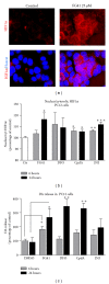
FG41 induces stabilization and nuclear translocation of HIF-1α and dopamine release in PC12 cells. (a) Immunocytochemical detection of HIF-1α in PC12 cells after 3 hours treatment with FG41 (25 and 50 μM). (b) Cellomics ArrayScan quantification of the nuclear to cytoplasmatic ratio of HIF-1α after 6 or 24 hours of treatment with FG41 or Cpd (both used at 25 μM). (d) DA release from PC12-cells induced by 6 or 24 hours of treatment with FG41 or CpdA (25 μM).

Similar articles

Cited by

References

    1. Kaelin WG, Jr., Ratcliffe PJ. Oxygen sensing by metazoans: the central role of the HIF hydroxylase pathway. Molecular Cell. 2008;30(4):393–402. - PubMed
    1. Epstein ACR, Gleadle JM, McNeill LA, et al. C. elegans EGL-9 and mammalian homologs define a family of dioxygenases that regulate HIF by prolyl hydroxylation. Cell. 2001;107(1):43–54. - PubMed
    1. Berra E, Benizri E, Ginouvès A, Volmat V, Roux D, Pouysségur J. HIF prolyl-hydroxylase 2 is the key oxygen sensor setting low steady-state levels of HIF-1α in normoxia. The EMBO Journal. 2003;22(16):4082–4090. - PMC - PubMed
    1. Sridharan V, Guichard J, Bailey RM, Kasiganesan H, Beeson C, Wright GL. The prolyl hydroxylase oxygen-sensing pathway is cytoprotective and allows maintenance of mitochondrial membrane potential during metabolic inhibition. American Journal of Physiology—Cell Physiology. 2007;292(2):C719–C728. - PubMed
    1. Sridharan V, Guichard J, Li CY, Muise-Helmericks R, Beeson CC, Wright GL. O2-sensing signal cascade: clamping of O2 respiration, reduced ATP utilization, and inducible fumarate respiration. American Journal of Physiology—Cell Physiology. 2008;295(1):C29–C37. - PMC - PubMed

LinkOut - more resources