. 2014:2014:851349.
doi: 10.1155/2014/851349. Epub 2014 May 21.

Atg6/UVRAG/Vps34-containing lipid kinase complex is required for receptor downregulation through endolysosomal degradation and epithelial polarity during Drosophila wing development

Affiliations

Atg6/UVRAG/Vps34-containing lipid kinase complex is required for receptor downregulation through endolysosomal degradation and epithelial polarity during Drosophila wing development

Péter Lőrincz et al. Biomed Res Int. 2014.

Abstract

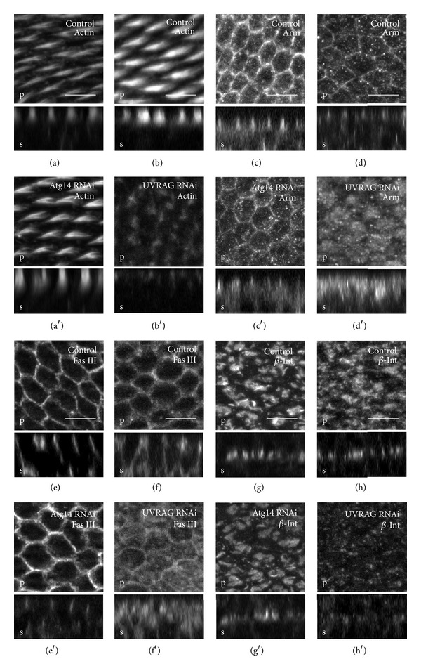
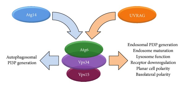
Atg6 (Beclin 1 in mammals) is a core component of the Vps34 PI3K (III) complex, which promotes multiple vesicle trafficking pathways. Atg6 and Vps34 form two distinct PI3K (III) complexes in yeast and mammalian cells, either with Atg14 or with UVRAG. The functions of these two complexes are not entirely clear, as both Atg14 and UVRAG have been suggested to regulate both endocytosis and autophagy. In this study, we performed a microscopic analysis of UVRAG, Atg14, or Atg6 loss-of-function cells in the developing Drosophila wing. Both autophagy and endocytosis are seriously impaired and defective endolysosomes accumulate upon loss of Atg6. We show that Atg6 is required for the downregulation of Notch and Wingless signaling pathways; thus it is essential for normal wing development. Moreover, the loss of Atg6 impairs cell polarity. Atg14 depletion results in autophagy defects with no effect on endocytosis or cell polarity, while the silencing of UVRAG phenocopies all but the autophagy defect of Atg6 depleted cells. Thus, our results indicate that the UVRAG-containing PI3K (III) complex is required for receptor downregulation through endolysosomal degradation and for the establishment of proper cell polarity in the developing wing, while the Atg14-containing complex is involved in autophagosome formation.


Figures

Figure 1
Figure 1
RT-PCR analysis of Atg6, UVRAG, or Atg14 transcripts from animals expressing transgenic RNAi constructs. Expression of Atg6, UVRAG, or Atg14 RNAi by tubGal4 strongly reduced the mRNA level of the corresponding genes. Interestingly, we found that the RNAi of Atg6 also decreased the mRNA level of Atg14.
Figure 2
Figure 2
Knockdown of Atg6 and UVRAG. (a) The PI3P marker GFP-2xFYVE localized to dots and vesicle-like structures in wild type pupal wings. (b), (d) In Atg6 or UVRAG depleted wings the GFP-2xFYVE became dispersed within the cytoplasm, indicating a failure in PI3P production. (c) In contrast to Atg6 or UVRAG RNAi wings the localization of GFP-2xFYVE did not differ from controls in Atg14 RNAi wings. (e) Quantification of (a)–(d). (f), (g) In control wings clathrin coated vesicle marker Clc-GFP showed punctate pattern which was unaffected by Atg6 RNAi. (h) Quantification of (f), (g). (i)–(w) Images from pupal wings expressing endosomal and lysosomal fluorescent markers controlled by a constitutive promoter (tubulin promoter for Rab-FP-s and Lamp1-GFP, cathD promoter for CathD-mCherry) and expressing RNAi constructs by enGal4. The region of RNAi is marked by the coexpression of RFP or GFP and the borderline of the region of RNAi is indicated with a dashed white line. (i)–(p) The number of structures positive for endosomal markers ((i): Rab4-YFP; (k): Rab5-CFP; (m): Rab7-YFP, except (o): Rab11-YFP) was significantly increased in Atg6 RNAi cells. (j), (l), (n), and (p) Quantification of (i), (k), (m), and (o), respectively. (q)–(t) The number of lysosomes marked by Lamp1-GFP or CathD-mCherry was significantly increased in Atg6 depleted cells. (r), (t) Quantification of (q), (s), respectively. (u) UVRAG RNAi resulted in the accumulation of Lamp1-GFP structures very similar to Atg6 RNAi. (v) Quantification of (u). (w) RNAi of Atg14 did not alter the number of Lamp1-GFP positive dots. (x) Quantification of (w). On images from (i) to (w), the intensity of the markers is enhanced by immunostaining the wings with an anti-GFP or anti-mCherry. Note the presence of RFP positive hemocytes under the epithelia on (i), (m), and (o) (marked by asterisks). On box plots, bars show the data ranging between the upper and lower quartiles; median of the signal covered areas is indicated as a horizontal black line within the box. Whiskers plot the smallest and largest observations, while dots and asterisks indicate outliers. NS means P > 0.05 and ∗∗∗ means P < 0.001. For details and exact P values of statistical analyses, see Table S3. For genotypes see Table S4. Scale bars represent 15 μm.
Figure 3
Figure 3
Electron microscopy of pupal wings. (a) Ultrastructure of control cells. (b) Depletion of Atg6 by RNAi resulted in the massive accumulation of enlarged multivesicular bodies (asterisks), dense multivesicular-body like structures (arrows), and multilamellar bodies (open arrows). (c) All of these structures could be found in Atg6 mutant pupal wings. (d) The depletion of Atg8a did not alter the ultrastructure of pupal wing cells. (e) Similar to Atg6, UVRAG RNAi also resulted in the accumulation of aberrant endolysosome-like structures (MVBs: arrows, MLBs: open arrows). (f) The depletion of Atg14 did not alter the ultrastructure of pupal wing cells. M: mitochondria, L: lipid droplet, and Ly: lysosome. For genotypes, see Table S4. Scale bars represent 1 μm.
Figure 4
Figure 4
Identification of the abnormal organelles of Atg6 RNAi cells. (a), (b) Immunogold labeling of Atg6 RNAi cells with anti-GFP antibody to detect Lamp1-GFP. MVBs, dMVBs, and MLBs were positive to Lamp1-GFP. (c) Enzymecitochemistry revealed small, dense acid phosphatase positive structures (open arrow) in wild type pupal wing cells. Based on the size and morphology, these structures were considered as primary lysosomes. All other organelles were devoid of reaction product. (d) Electron-lucent MVBs in Atg6 depleted cells never contained acid phosphatase reaction product but were often found in the vicinity of acid phosphatase positive primary lysosomes (open arrow). (e) A dense multivesicular body-like structure (dMVB) showing acid phosphatase reaction product in an Atg6 RNAi pupal wing cell. (f) A multilamellar body (MLB) in Atg6 depleted cell positive to acid phosphatase. (g), (h) DAB-staining of pupal wings expressing plasma membrane localized horseradish peroxidase enzyme (HRP-CD2). (g) In control cells plasma membrane showed a prominent DAB staining (arrowheads), just as organelles with plasma membrane origin, such as early endosomes (EE), multivesicular bodies (MVB), and secondary lysosomes (Ly) as well. (h) The accumulated organelles in Atg6 depleted cells: the multivesicular bodies (arrows) and multilamellar bodies (open arrows) were all positive to DAB. M: mitochondria, MVB: multivesicular body, dMVB: dense multivesicular body-like structure, MLB: multilamellar body, N: nucleus, Ns: nucleolus, and Ly: lysosome. For genotypes, see Table S4. Scale bars represent 200 nm in (a)–(f) and 500 nm in (g), (h).
Figure 5
Figure 5
Atg6 and Atg14 are required for autophagy in contrast to UVRAG. (a)–(d) Larval wing discs expressing RNAi with transgenic Myc along with the autophagy marker mCherry-Atg8 in the patched (ptc) domain using ptcGal4. (a) In controls the expression of Myc induces massive autophagy in the ptc domain. (b) Atg6 or (c) Atg14 RNAi inhibited Myc-induced autophagy as the pattern of mCherry-Atg8 became dispersed in the citoplasm, whereas (d) UVRAG RNAi did not alter the pattern of the autophagy marker. (e), (g), and (i) Pupal wings expressing RNAi constructs by enGal4, immunostained against the selective autophagic cargo protein p62. (e) Atg6 or (g) Atg14 RNAi resulted in the massive accumulation of p62 aggregates. (i) UVRAG RNAi cells did not accumulate p62 aggregates. (f), (h), and (j) Quantification of (e), (g), and (i), respectively. On box plots, bars show the data ranging between the upper and lower quartiles; median of the signal covered areas is indicated as a horizontal black line within the box. Whiskers plot the smallest and largest observations, while dots and asterisks indicate outliers. NS means P > 0.05 and ∗∗∗ means P < 0.001. For details and exact P values of statistical analyses, see Table S3. For genotypes, see Table S4. Scale bars represent 25 μm in (a)–(d) and 15 μm in (e), (g), and (i).
Figure 6
Figure 6
Impaired endocytic degradation of Notch in Atg6 depleted cells. (a) In 3rd instar larval wing discs Atg6 null mutant clones (marked by the absence of GFP, asterisks) accumulate the extracellular domain of Notch (NECD). (b) Quantification of (a). (c), (e), and (g) Imaginal wing discs expressing dsRNA (Atg6, Atg14) or siRNA (UVRAG) by enGal4 were immunostained against NECD. The region of RNAi is marked by the coexpression of GFP and the borderline of the region is indicated with a dashed white line. Immunostain revealed that NECD accumulated in small, numerous puncta in the case of (c) Atg6 RNAi and (e) UVRAG RNAi, (g) but not in Atg14 RNAi. (d), (f), and (h) Quantification of (c), (e), and (g), respectively. (i) Live trafficking assay for Notch in cultured wing imaginal discs. Atg6 RNAi region is marked by the coexpression of the lysosome marker Lamp1-GFP. In controls, surface-bound Notch was internalized then degraded after 3 h. In contrast, in Atg6 depleted cells Notch was internalized normally but trapped in vesicular structures even at 3 h of chasing. Arrows indicate Notch signal colocalizing with Lamp1-GFP. On box plots, bars show the data ranging between the upper and lower quartiles; median of the signal covered areas is indicated as a horizontal black line within the box. Whiskers plot the smallest and largest observations, while dots and asterisks indicate outliers. NS means P > 0.05 and ∗∗∗ means P < 0.001. For details and exact P values of statistical analyses, see Table S3. For genotypes, see Table S4. Scale bars represent 50 μm.
Figure 7
Figure 7
Enhanced Notch signaling in Atg6 RNAi and UVRAG RNAi cells. Larval wing discs expressing RNAi with or without transgenic Notch. UAS-myrRFP was used to mark enGal4-specific region. Notch response element- (NRE-) EGFP was used to show Notch transcriptional activity. (a) Atg6 RNAi and (b) Notch overexpression lead to enhanced Notch activity. (c) This effect was further increased as coexpression of Atg6 RNAi and Notch strongly enhanced the reporter expression. (d) UVRAG enhanced Notch transcriptional activity similarly to Atg6 RNAi (e) RNAi of Atg14 did not alter the reporter expression. (f) Control disc. Histograms show the intensity profiles of the NRE-GFP expression in the enGal4-specific regions. Note that the peaks are broader in panels (a)–(d), compared to panels (e), (f). For genotypes, see Table S4. Scale bars represent 50 μm.
Figure 8
Figure 8
Impaired endocytic degradation of Wnt in Atg6 or UVRAG depleted cells. Imaginal wing discs expressing RNAi by enGal4 were immunostained against Wnt. The region of RNAi is marked by the coexpression of GFP and the borderline of the region is indicated with a dashed white line (a), (b) RNAi of Atg6 and UVRAG resulted in the accumulation of Wnt in small, numerous intracellular puncta. (c) RNAi of Atg14 did not affect Wnt localization. For genotypes, see Table S4. Scale bars represent 50 μm.
Figure 9
Figure 9
Atg6 and the other members of PI3K (III) are required for normal wing development: (a), (f) 1-day-old wild type flies. (b) Newly hatched Atg6 RNAi animals developed serious malformations, blisters in their wings. (c), (d) In 1-day-old Atg6 RNAi animals, the blisters had become collapsed; thus the wings were creased and heavily distorted. (g), (h) The RNAi of UVRAG completely mimiced the effect of Atg6 RNAi. (e), (f) Positive control animals: Vps34 RNAi and Vps15 RNAi flies, respectively. (j) Unlike the other members of PI3K (III) complex, the RNAi of Atg14 causes a vestigial-like morphology rather than blistering or creasing. (k) The Atg6 null mutant wings are heavily distorted and creased, (l) compared to the control wings lacking the mutation or (m) expressing an Atg6 transgene. 1 h: 1 hour; 1 d: 1 day after emerging from puparia, respectively. For genotypes, see Table S4.
Figure 10
Figure 10
Polarity and membrane protein localization defects of Atg6-knockdown pupal wings. Semiconfocal immunodetection showing XY (marked by a letter p (plan view) at the bottom left corners) and XZ sections (marked by a letter s (side view) at the bottom left corners) in the plane of pupal wings at 32 h APF. Regions of controls are shown in boxes marked by a single character at the upper left side, whereas the regions where dsRNA for Atg6 was expressed by en-Gal4 driver are shown in boxes marked by a character at the upper left side with an apostrophe. (a) Low magnification image of a pupal wing with rhodamine-phalloidin stain showing the regions from where the high magnification images were taken. The expression of the dsRNA by enGal4 is restricted to the posterior compartment of the wing (marked by the coexpression of GFP), and anterior side serves as control. Small boxes surround areas of images (b) and (b′) in which rhodamine-phalloidin stain reveals that control wing hairs are well-developed and very regularly oriented towards the distal end of the wing. In contrast the wing hairs are poorly developed or completely absent in the regions of the RNAi. (c)–(e) In controls Fmi, Arm, and DE-Cad show a very pronounced zonula adherens (ZA) localization; furthermore Fmi shows a very regular planar cell polarity pattern as well, whereas all of them show intracellular punctuation and irregular localization in the Atg6 depleted wing regions. (f), (g) Septate junction proteins Fas III and Dlg show very typical lateral localizations as Dlg is mainly localized at the apical regions of septate junctions (SJ), whilst the domain of Fas III is expanded throughout the whole SJ. In contrast, in Atg6 RNAi cells the amounts of these proteins lowered and both show less pronounced SJ localization. (h) At this developmental stage the prospective wing intervein cells form large, β-integrin (β-int) containing basal junctions between the two epithelial layers of the wing, but this process is dramatically blocked when the level of Atg6 is reduced and wing cells seem to be unable to develop these structures. For genotype, see Table S4. Scale bars represent (a) 10 μm and (b)–(h) 5 μm.
Figure 11
Figure 11
RNAi of UVRAG but Atg14 alters cell polarity and membrane protein localization. Semiconfocal immunodetection showing XY (marked by a letter p (plan view) at the bottom left corners) and XZ sections (marked by a letter s (side view) at the bottom left corners) in the plane of pupal wings at 32 h APF. Regions of controls are shown in boxes marked by a single character at the upper left side, whereas the regions where dsRNA for Atg6 was expressed by en-Gal4 driver are shown in boxes marked by a character at the upper left side with an apostrophe. (a), (c), (e), and (g) Immunodetection reveals no obvious differences of the pattern of wing hairs and the localization of Fas III, Arm, and β-int between control and Atg14 depleted regions of pupal wings. (b), (d), (f), and (h) In contrast to Atg14, the RNAi of UVRAG dramatically alters wing hair development and the localization of membrane proteins (Fas III, Arm, and β-int) and these phenotypes are very similar to those induced by Atg6 RNAi as was shown in Figure 2. For genotypes, see Table S4. Scale bars represent 5 μm.
Figure 12
Figure 12
Schematic representation summarizing the multiple roles of the two PI3K (III) complexes in Drosophila. See text for details.

Similar articles

Cited by

References

    1. Mizushima N, Yoshimori T, Ohsumi Y. The role of atg proteins in autophagosome formation. Annual Review of Cell and Developmental Biology. 2011;27:107–132. - PubMed
    1. Érdi B, Nagy P, Zvara Á, et al. Loss of the starvation-induced gene Rack1 leads to glycogen deficiency and impaired autophagic responses in Drosophila. Autophagy. 2012;8:1124–1135. - PMC - PubMed
    1. Itakura E, Kishi-Itakura C, Mizushima N. The hairpin-type tail-anchored SNARE syntaxin 17 targets to autophagosomes for fusion with endosomes/lysosomes. Cell. 2012;151:1256–1269. - PubMed
    1. Takáts S, Nagy P, Varga Á, et al. Autophagosomal syntaxin17-dependent lysosomal degradation maintains neuronal function in Drosophila. The Journal of Cell Biology. 2013;201:531–539. - PMC - PubMed
    1. Hegedűs K, Takáts S, Kovács AL, Juhász G. Evolutionarily conserved role and physiological relevance of a STX17/Syx17 (syntaxin 17)-containing SNARE complex in autophagosome fusion with endosomes and lysosomes. Autophagy. 2013;9:1642–1646. - PubMed

Publication types

MeSH terms

LinkOut - more resources