. 2014 Jul 9:5:4366.
doi: 10.1038/ncomms5366.

G1-arrested newborn cells are the predominant infectious form of the pathogen Brucella abortus

Affiliations

G1-arrested newborn cells are the predominant infectious form of the pathogen Brucella abortus

Michaël Deghelt et al. Nat Commun. .

Abstract

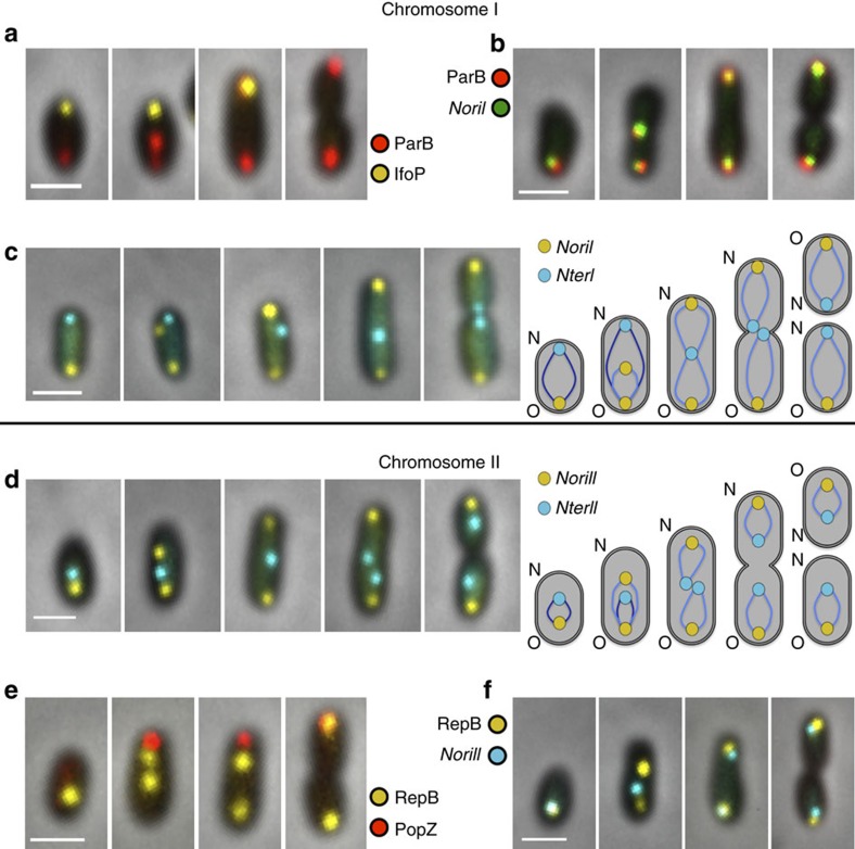
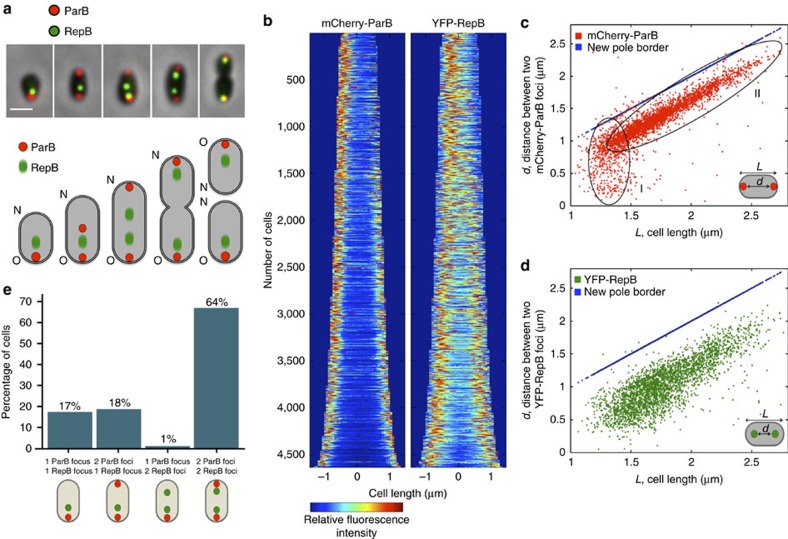
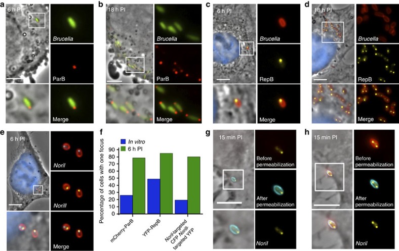
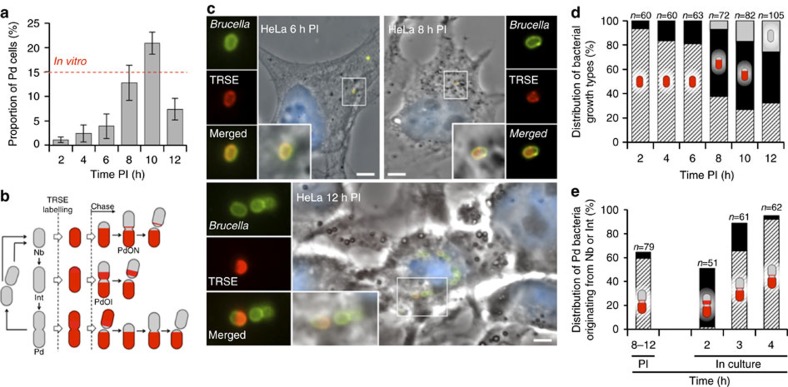
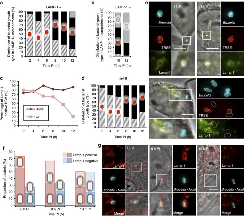
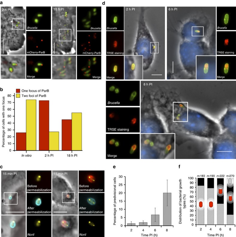
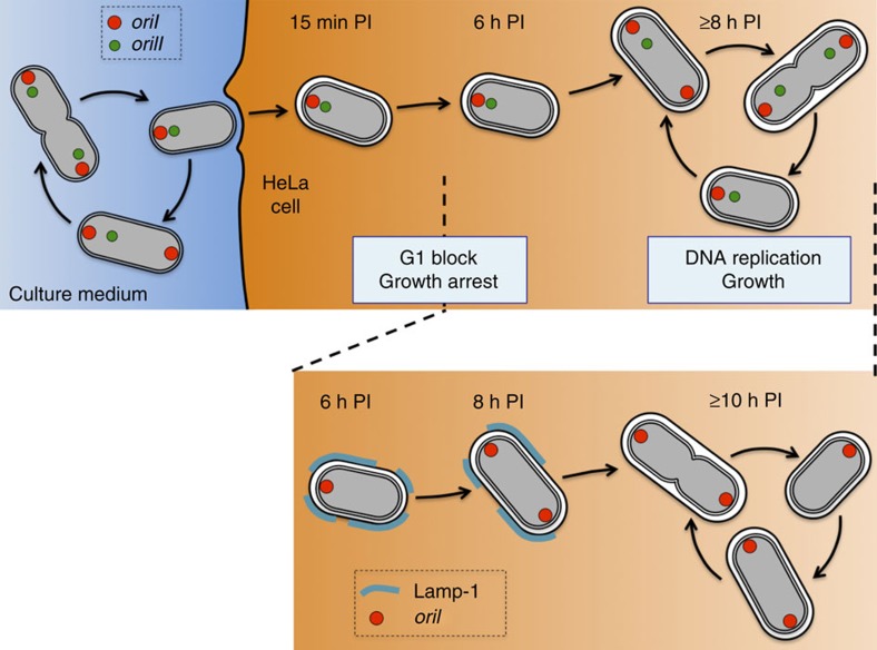
Several intracellular pathogens, such as Brucella abortus, display a biphasic infection process starting with a non-proliferative stage of unclear nature. Here, we study the cell cycle of B. abortus at the single-cell level, in culture and during infection of HeLa cells and macrophages. The localization of segregation and replication loci of the two bacterial chromosomes indicates that, immediately after being engulfed by host-cell endocytic vacuoles, most bacterial cells are newborn. These bacterial cells do not initiate DNA replication for the next 4 to 6 h, indicating a G1 arrest. Moreover, growth is completely stopped during that time, reflecting a global cell cycle block. Growth and DNA replication resume later, although bacteria still reside within endosomal-like compartments. We hypothesize that the predominance of G1-arrested bacteria in the infectious population, and the bacterial cell cycle arrest following internalization, may constitute a widespread strategy among intracellular pathogens to colonize new proliferation niches.


Figures

Figure 1
Figure 1. Localization patterns of segregation markers and specific genomic regions of both B. abortus chromosomes.
(a) Representative cells producing mCherry-ParB (red) and IfoP-YFP (yellow, new pole marker, see Supplementary Fig. 4). The second picture shows a probable recent event of ParB duplication. (b) Colocalization between mCherry-ParB (red) and NoriI-targeted YFP (green) in representative cells. (c) Typical NoriI-targeted YFP (yellow) and NterI-targeted CFP (blue) localization patterns and schematic representation of chrI orientation along the bacterial cell cycle. Polar NterI region was never found when duplicated NoriI is bipolar. O represents the old pole and N the new pole locations. (d) Typical NoriII-targeted YFP (yellow) and NterII-targeted CFP (blue) localization patterns and schematic representation of chrII orientation along the bacterial cell cycle. (e) Representative cells producing YFP-RepB (yellow) and PopZ-mCherry (red) fusions (PopZ-mCherry is a new pole marker, see Supplementary Fig. 7). (f) Partial colocalization between YFP-RepB (yellow) and NoriII-targeted CFP (blue). Larger fields of the different localization patterns are available in Supplementary Fig. 3a for mCherry-ParB and IfoP-YFP, in Supplementary Fig. 3d for mCherry-ParB and NoriI-targeted YFP, in Supplementary Fig. 6a for NoriII-YFP and NterII-CFP, in Supplementary Fig. 6d for YFP-RepB and NoriII-CFP. Scale bars, 1 μm.
Figure 2
Figure 2. Distinct localization patterns of the two replication origins and systematic delay of replication initiation of chrII.
(a) Typical localization patterns of cells producing mCherry-ParB (red) and YFP-RepB (green) fusions and schematic representation of their positions. Fuzzy green dots represent the intracellular mobility of YFP-RepB. A larger field of view is available at Supplementary Fig. 9c. O represents the old pole and N the new pole locations. Scale bar, 1 μm. (b) Demographic representation of mCherry-ParB and YFP-RepB fusions (n=4,636 cells). Cells are sorted according to their length (small cells are on the top), and in each cell the fluorescence intensity of mCherry-ParB or YFP-RepB is represented by a colour code, the low intensity corresponding to dark blue, while high intensity is shown in red (cf) Representation of the distance between two mCherry-ParB or two YFP-RepB foci according to the cell length. (c) Distance between two mCherry-ParB foci can be classified in two specific classes (I and II) suggesting a two-step segregation mechanism. (d) Distance between two YFP-RepB foci display a single progressive separation process. (e) Proportion of cells for different numbers of mCherry-ParB (red) and YFP-RepB (yellow) spots (n=4,636 cells). Almost no bacteria displaying two RepB foci but only one ParB spot were detected suggesting that replication initiation of chrI origin occurs before replication of chrII.
Figure 3
Figure 3. Single foci of mCherry-ParB, YFP-RepB, CFP-labelled NoriI and YFP-labelled NoriII fusions are predominant at 6 h PI.
(a,b) Bacteria producing mCherry-ParB (red) fusion and diffuse GFP (green) at (a) 6 h and (b) 18 h PI. At 6 h PI, most bacteria showed (79%) one mCherry-ParB focus. (c,d) B. abortus (red) producing YFP-RepB (yellow) fusion at 6 h (c) and 18 h (d) PI respectively. At 6 h PI, most bacteria showed (85%) one YFP-RepB focus. (e) B. abortus (red) with CFP-labelled NoriI (cyan) and YFP-labelled NoriII (yellow) at 6 h PI. Most bacteria showed (80%) one focus for both fusions. In c to e, nuclei are stained with 4',6-diamidino-2-phenylindole (blue). (f) Comparison of the percentage of cells displaying one fluorescent focus for mCherry-ParB, YFP-RepB, CFP-labelled NoriI and YFP-labelled NoriII respectively in vitro (grey) and 6 h PI (black) conditions. (g,h) B. abortus YFP-labelled NoriI at 15 min PI after performing a dual labelling. Intracellular bacteria (g) are not labelled with antibodies used before permeabilization (red), while extracellular bacteria (h) are recognized by antibodies used before and after permeabilization (red and cyan, respectively). One or two intracellular bacteria were found in each infected cell, and most of these bacteria displayed only one focus of YFP-labelled NoriI (73%). Scale bars, 5 μm.
Figure 4
Figure 4. Growth characterization of B. abortus during HeLa cells infection.
(a) Proportion of Pd bacterial type at different times PI. The red dashed line represents the proportion of Pd bacteria detected in culture. Error bars represent s.d. The number of bacteria counted at 2, 4, 6, 8, 10 and 12 h PI is 357, 359, 372, 360, 376 and 385, respectively. One hundred and sixty-eight bacteria were counted in culture. (b) Schematic representation of the evolution of unipolarly growing cell types after a TRSE labelling (red areas). Nb stands for newborn, Int for intermediate and Pd for predivisional. PdON stands for predivisional originating from a newborn cell (weak TRSE labelling in the newly generated part) and PdOI for predivisional originating from an intermediate cell (partial TRSE labelling in the newly generated part). (c) Representative IF images of TRSE labelled B. abortus after 6, 8 and 12 h of infection in HeLa cells. The bacteria were labelled in green. Scale bars, 1 μm. (d) Distribution of the three observed types of TRSE-labelled bacteria; completely labelled (hatched), partially labelled (black) and non-labelled (grey), at different times PI. Up to 6 h PI, the majority of bacteria show no sign of growth. The number of counted intracellular bacteria (n) is indicated for each time PI. (e) Growth resumption of TRSE-labelled bacteria at for 2, 3 or 4 h in culture or used for infection and observed after 8 to 12 h PI in HeLa cells. The proportion of PdON cells (dashed lines) or PdOI cells (black) was determined. After 8 to 12 h PI, the majority of the growing Pd cell types are originating from the Nb type (PdON). In culture, a majority of PdOI are observed before a complete generation time (that is, 3 h) suggesting that intermediate cells were predominant compared with newborns in culture.
Figure 5
Figure 5. Bacterial cell cycle resumes while B. abortus is still associated with Lamp-1-positive compartments.
(a) Distribution of the three observed types of TRSE-labelled bacteria; completely labelled (hatched), partially labelled (black) and unlabelled (grey), at different times PI in Lamp-1-positive compartments. At 8 h PI, a large proportion of bacteria residing in Lamp-1-positive compartments started to grow. Fourty-three to 103 bacteria are counted for each time PI. (b) Distribution of the bacterial growth types in Lamp-1-negative compartments at 10 and 12 h PI. The majority of newly generated bacteria (unlabelled by TRSE) are found in Lamp-1-negative compartments. (c) Proportion of Lamp-1-positive Brucella-containing vacuoles (BCV) along the trafficking of the wild-type (wt) strain and the ΔvirB strain. While the ΔvirB strain-containing vacuoles retained Lamp-1 labelling, the wt strain lost Lamp-1 staining progressively, with a marked decrease at 10 and 12 h PI. (d) Distribution of the three observed types of TRSE-labelled bacteria in Lamp-1-positive compartments for the ΔvirB strain. A majority of partially labelled bacteria are observed from 8 h PI, while no newly generated bacteria are observed at any time PI, in agreement with the previously reported absence of intracellular proliferation of the virB mutant. 43 to 141 bacteria are counted for each time PI. (e) TRSE-labelled B. abortus strain observed at 6, 8 and 12 h PI in HeLa cells labelled by anti-Lamp-1 staining. (f) Distribution of the foci number of YFP-labelled NoriI in Lamp-1 stained vacuoles in HeLa cells. In each type of vacuole (Lamp-1-positive or Lamp-1-negative), the proportion of bacteria with two foci of YFP-labelled NoriI is shown with a light colour. The proportion of bacteria displaying two NoriI foci increases in Lamp-1-positive compartments at 8 h PI and in Lamp-1-negative compartments at 10 h PI. The total number of counted bacteria was 223, 228 and 277 for 6, 8 and 10 h PI respectively. (g) B. abortus YFP-labelled NoriI strain observed at 6, 8 and 10 h PI in HeLa cells labelled by anti-Lamp-1 staining. Scale bars represent 5 μm.
Figure 6
Figure 6. G1 phase block and growth arrest of B. abortus during the non-proliferative phase of the infection in RAW264.7 macrophages.
(a,b) Strain producing mCherry-ParB (red) and diffuse GFP (green) in RAW264.7 macrophages at 2 and 18 h PI. At 2 h PI, 72.9% of the bacteria display only one mCherry-ParB focus. (c) B. abortus YFP-labelled NoriI at 15 min PI after performing a dual labelling to discriminate between extracellular bacteria (red and cyan) from intracellular bacteria (cyan only). Only one or two intracellular bacteria were found per infected cell, these bacteria mainly displayed only one focus of YFP-labelled NoriI (77%). (d) TRSE-labelled B. abortus strain observed at 2, 6 and 8 h PI in RAW264.7 macrophages. Scale bars in (a), (c) and (d) represent 5 μm. (e) Proportion of predivisional (Pd) B. abortus at 2, 4, 6 and 8 h PI (n=183, 190, 222 and 270, respectively). This proportion is higher at 8 h PI. Error bars represent s.d. (f) Distribution of the three observed types of TRSE-labelled bacteria; completely labelled (hatched), partially labelled (black) and non-labelled (grey), at different times PI in RAW264.7 macrophages. Up to 4 h PI, the majority of bacteria show no sign of growth. The number of counted intracellular bacteria (n) is indicated for each time PI.
Figure 7
Figure 7. Model of the B. abortus cell cycle outside and inside host cells.
During the B. abortus cell cycle, replication of chrI is initiated before chrII. Newborn B. abortus (in G1) are proposed to be the predominantly invasive cell type for host cells. Indeed, between 15 min and 6 h PI, that is, the non-proliferative stage of the infection, bacteria are found as non-growing cells blocked before the replication initiation of the two chromosomes. Growth and chromosomal replication are reiniated in Lamp-1-positive compartments at 8 h PI (lower panel), and cell division is completed in Lamp-1-negative compartments, where bacteria subsequently proliferate.

Comment in

Similar articles

Cited by

References

    1. Salcedo S. P. & Holden D. W. Bacterial interactions with the eukaryotic secretory pathway. Curr. Opin. Microbiol. 8, 92–98 (2005). - PubMed
    1. Moreno E. & Moriyon I. Prokaryotes 5, 315–456Springer (2006).
    1. Comerci D. J., Martinez-Lorenzo M. J., Sieira R., Gorvel J. P. & Ugalde R. A. Essential role of the VirB machinery in the maturation of the Brucella abortus-containing vacuole. Cell. Microbiol. 3, 159–168 (2001). - PubMed
    1. Celli J. et al. Brucella evades macrophage killing via VirB-dependent sustained interactions with the endoplasmic reticulum. J. Exp. Med. 198, 545–556 (2003). - PMC - PubMed
    1. Pizarro-Cerda J. et al. Brucella abortus transits through the autophagic pathway and replicates in the endoplasmic reticulum of nonprofessional phagocytes. Infect. Immun. 66, 5711–5724 (1998). - PMC - PubMed

Publication types

LinkOut - more resources