. 2014 Jul 8;19(7):9876-92.
doi: 10.3390/molecules19079876.

Fluorescent lectins for local in vivo visualization of peripheral nerves

Affiliations

Fluorescent lectins for local in vivo visualization of peripheral nerves

Gijs Hendrik KleinJan et al. Molecules. .

Abstract

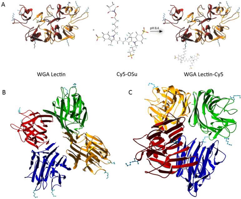
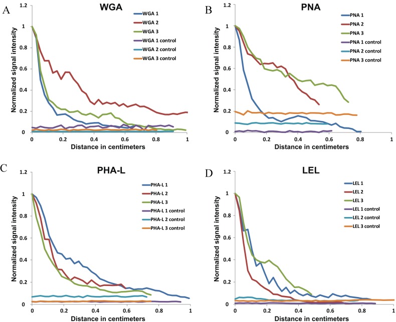
Damage to peripheral nerves caused during a surgical intervention often results in function loss. Fluorescence imaging has the potential to improve intraoperative identification and preservation of these structures. However, only very few nerve targeting agents are available. This study describes the in vivo nerve staining capabilities of locally administered fluorescent lectin-analogues. To this end WGA, PNA, PHA-L and LEL were functionalized with Cy5 (λex max 640 nm; λem max 680 nm). Transfer of these imaging agents along the sciatic nerve was evaluated in Thy1-YFP mice (n = 12) after intramuscular injection. Migration from the injection site was assessed in vivo using a laboratory fluorescence scanner and ex vivo via fluorescence confocal microscopy. All four lectins showed retrograde movement and staining of the epineurium with a signal-to-muscle ratio of around two. On average, the longest transfer distance was obtained with WGA-Cy5 (0.95 cm). Since WGA also gave minimal uptake in the lymphatic system, this lectin type revealed the highest potential as a migration imaging agent to visualize nerves.


Conflict of interest statement

The authors declare no conflict of interest.

Figures

Figure 1
Figure 1
Crystal structures and method of labeling. (A) WGA, consisting of two subunits, with four readily available lysines per subunit, covalently attached to sulphonated Cy5-OSu. (B) PHA-L, consisting of four subunits, with two readily available lysines per subunit. (C) PNA, consisting of four subunits forming a tetrahedral structure, with two readily available lysines per subunit. To the best of our knowledge, the crystal structure for LEL (tomato lectin) is unknown.
Figure 2
Figure 2
Cy5/Lectin labeling ratio per lectin.
Figure 3
Figure 3
In vivo distribution of locally injected lectins (A) Schematic representation of the local injection of Cy5-lectins and the corresponding migration path. The injection site (IS) and sciatic nerve (N) are depicted in blue and green respectively. The lymphatic tract leading from the IS to the lymph node (LN) is shown in red. (B) Illustration of the YFP signal in the nerves (green) which served as control for the localization of the nerves. Representative images of the injection site before (C) and after (D) removal of the nerve, and excised nerves (E). In each of these pictures, yellow arrows -the injection site; purple arrows -fluorescence signal from injection site and nerve, red arrows -the control side nerve and light blue arrows -the control nerve.
Figure 4
Figure 4
Migration curves. Normalized curves of (A) WGA, (B) PNA, (C) PHA-L and (D) LEL show the migration of the lectins along the individual nerves. On the y-axis the normalized intensity signal is depicted, on the x-axis the migration distance (cm).
Figure 5
Figure 5
Schematic overview peripheral nerve wherein Cy5/ WGA lectin binds to PG’s located on the epineurium and extracellular matrix.
Figure 6
Figure 6
Binding mode after in vivo local administration. The fluorescence signal in nerves from Thy-1 YFP mice (YFP in green and Cy5 in red) was traced from (A) the injection site to (B) the middle and (C) the proximal side of the nerve. In all cases, staining of the epineurium was observed with a decrease in signal when the distance from the injection site increased.
Figure 7
Figure 7
Ex vivo incubation. Ex vivo incubation confirmed the in vivo localization of staining. (YFP in green and Cy5 in red)

References

    1. Mauroy B., Demondion X., Drizenko A., Goullet E., Bonnal J.L., Biserte J., Abbou C. The inferior hypogastric plexus (pelvic plexus): Its importance in neural preservation techniques. Surg. Radiol. Anat. 2003;25:6–15. doi: 10.1007/s00276-002-0083-9. - DOI - PubMed
    1. Stelzner S., Holm T., Moran B.J., Heald R.J., Witzigmann H., Zorenkov D., Wedel T. Deep pelvic anatomy revisited for a description of crucial steps in extralevator abdominoperineal excision for rectal cancer. Dis. Colon. Rectum. 2011;54:947–957. doi: 10.1097/DCR.0b013e31821c4bac. - DOI - PubMed
    1. Tewari A.K., Srivastava A., Huang M.W., Robinson B.D., Shevchuk M.M., Durand M., Sooriakumaran P., Grover S., Yadav R., Mishra N., et al. Anatomical grades of nerve sparing: A risk-stratified approach to neural-hammock sparing during robot-assisted radical prostatectomy (RARP) BJU Int. 2011;108:984–992. doi: 10.1111/j.1464-410X.2011.10565.x. - DOI - PubMed
    1. Pfister B.J., Gordon T., Loverde J.R., Kochar A.S., Mackinnon S.E., Cullen D.K. Biomedical engineering strategies for peripheral nerve repair: Surgical applications, state of the art, and future challenges. Crit. Rev. Biomed. Eng. 2011;39:81–124. doi: 10.1615/CritRevBiomedEng.v39.i2.20. - DOI - PubMed
    1. Myssiorek D. Recurrent laryngeal nerve paralysis: Anatomy and etiology. Otolaryngol. Clin. North. Am. 2004;37:25–44. doi: 10.1016/S0030-6665(03)00172-5. - DOI - PubMed

Publication types

LinkOut - more resources