. 2014 Sep;10(9):1549-64.
doi: 10.4161/auto.29329. Epub 2014 Jun 30.

The membrane peroxin PEX3 induces peroxisome-ubiquitination-linked pexophagy

Affiliations

The membrane peroxin PEX3 induces peroxisome-ubiquitination-linked pexophagy

Shun-ichi Yamashita et al. Autophagy. 2014 Sep.

Abstract

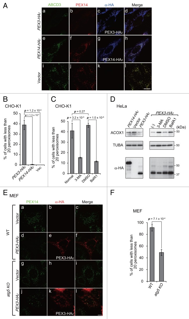
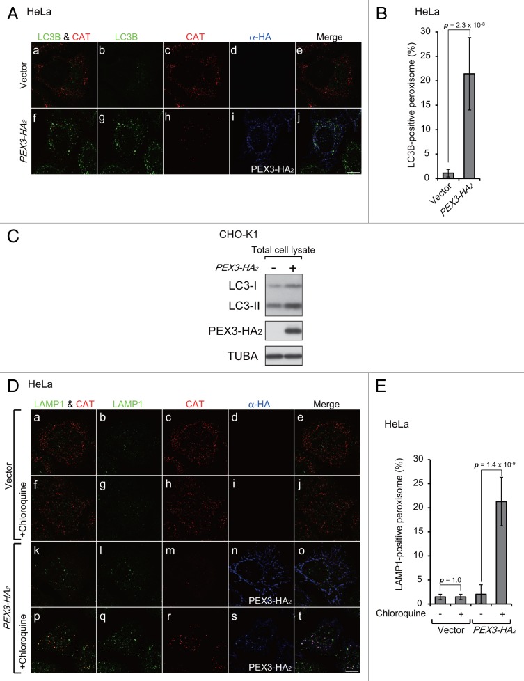
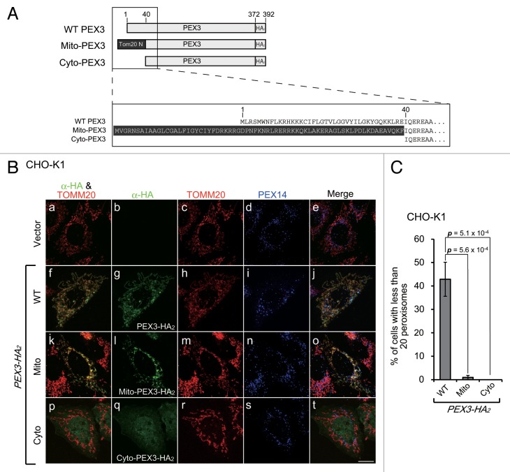
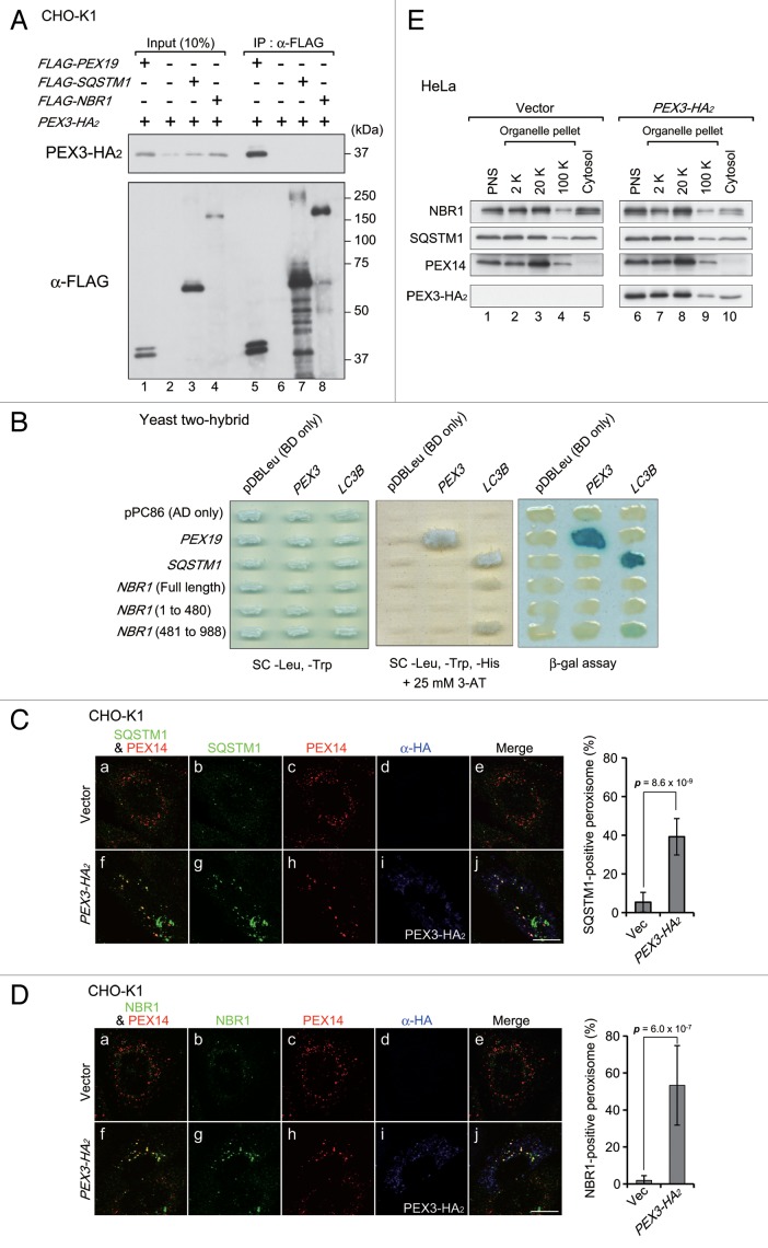
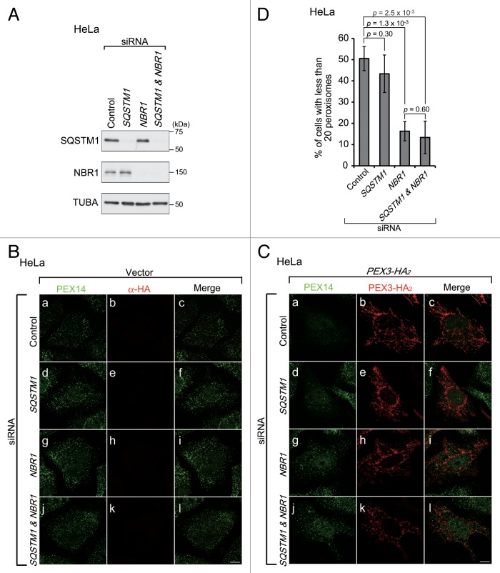
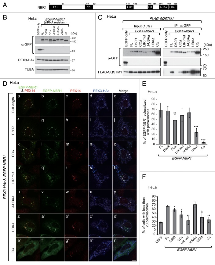
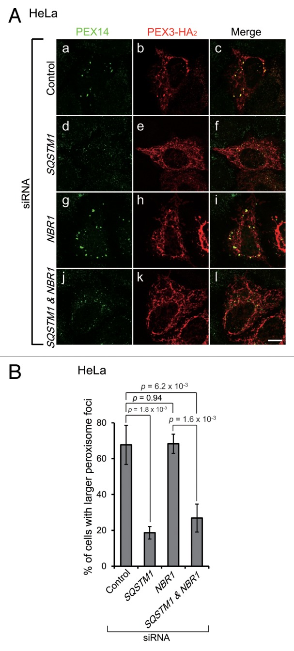
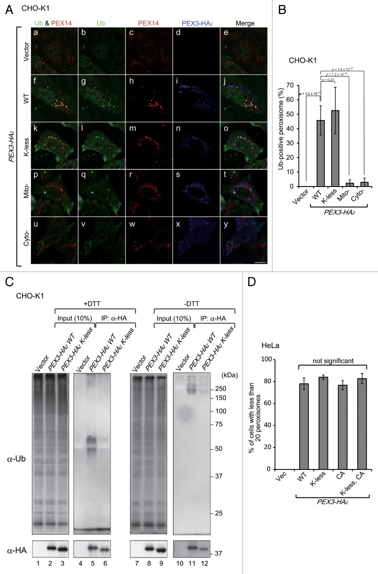
Peroxisomes are degraded by a selective type of autophagy known as pexophagy. Several different types of pexophagy have been reported in mammalian cells. However, the mechanisms underlying how peroxisomes are recognized by autophagy-related machinery remain elusive. PEX3 is a peroxisomal membrane protein (PMP) that functions in the import of PMPs into the peroxisomal membrane and has been shown to interact with pexophagic receptor proteins during pexophagy in yeast. Thus, PEX3 is important not only for peroxisome biogenesis, but also for peroxisome degradation. However, whether PEX3 is involved in the degradation of peroxisomes in mammalian cells is unclear. Here, we report that high levels of PEX3 expression induce pexophagy. In PEX3-loaded cells, peroxisomes are ubiquitinated, clustered, and degraded in lysosomes. Peroxisome targeting of PEX3 is essential for the initial step of this degradation pathway. The degradation of peroxisomes is inhibited by treatment with autophagy inhibitors or siRNA against NBR1, which encodes an autophagic receptor protein. These results indicate that ubiquitin- and NBR1-mediated pexophagy is induced by increased expression of PEX3 in mammalian cells. In addition, another autophagic receptor protein, SQSTM1/p62, is required only for the clustering of peroxisomes. Expression of a PEX3 mutant with substitution of all lysine and cysteine residues by arginine and alanine, respectively, also induces peroxisome ubiquitination and degradation, hence suggesting that ubiquitination of PEX3 is dispensable for pexophagy and an endogenous, unidentified peroxisomal protein is ubiquitinated on the peroxisomal membrane.

Keywords: NBR1; PEX3; SQSTM1/p62; autophagy; peroxisomal membrane protein; peroxisome; pexophagy; ubiquitin.


Figures

None
Figure 1. PEX3 overexpression induces pexophagy. (A) CHO-K1 cells were transfected with PEX3-HA2 (a–d), PEX3-HA2 (e–h) and empty vector (i and j), as indicated. After 24 h, the cells were fixed and immunostained with antibodies against ABCD3/PMP70 (a, e, and i), PEX14 (b, f, and j), and HA (c, g, and k). Merged views are shown (d, h, and l). (B and C) The percentage of cells showing fewer than 20 peroxisomes was calculated from 50 cells transfected with PEX3-HA2, PEX14-HA2 or empty vector alone as shown in (A, a, e, and i) (B) and those transfected with PEX3-HA2 in the presence of autophagy inhibitors (C). Data are presented as the mean ± SD of 3 replicates. 3-MA: 3-methyladenine (10 mM); BafA1: bafilomycinA1 (100 nM). (D) HeLa cells were transfected with PEX3-HA2 and PEX14-HA2 alone in the absence (-) or presence of autophagy inhibitors. After 24 h, the cells were lysed with SDS-PAGE sample buffer and analyzed by SDS-PAGE and immunoblotting with antibodies against ACOX1 (acyl-CoA oxidase), a peroxisomal matrix protein, and TUBA/α-tubulin for a loading control. (E) WT MEF (a–f) and atg5 KO MEFs (g–l) were transfected with PEX3-HA2 and immunostained with antibodies against PEX14 (a, d, g, and j) and HA (b, e, h, and k). (F) The percentage of cells showing less than 20 peroxisomes was calculated as in (B and C). Scale bars: 10 μm.
None
Figure 2. Peroxisomes are recruited into lysosomes upon PEX3 overexpression. (A) HeLa cells were transfected with empty vector (a–e) and PEX3-HA2 (f–j). After 12 h, the cells were fixed and immunostained with antibodies against LC3B (b and g), CAT (catalase) (c and h), and HA (d and i). Merged views of LC3B and CAT are shown in (a and f). (B) The percentage of peroxisome signals colocalized with LC3B was calculated by Metamorph version 7.6 software from 10 cells. (C) CHO-K1 cells transfected with empty vector and PEX3-HA2 were lysed and analyzed by SDS-PAGE and immunoblotting with antibodies against LC3 (upper panel), HA (middle panel), and TUBA (bottom panel). (D) HeLa cells were transfected with empty vector (a–j) and PEX3-HA2 (k–t) in the presence of 20 μM chloroquine, a lysosome inhibitor (f–j and p–t). After 24 h, the cells were fixed and immunostained with antibodies against LAMP1 (b, g, l, and q), CAT (c, h, m, and r) and HA (d, i, n, and s). Merged views of immunostained peroxisomes and LAMP1 are shown in (a, f, k, and p). (E) The percentage of peroxisome signals colocalized with LAMP1 was calculated using Metamorph version 7.6 software from 10 cells. Scale bars: 10 μm.
None
Figure 3. Peroxisomal membrane targeting of Pex3 is essential for the induction of pexophagy. (A) Schematic representation and alignment of N-terminal amino acid sequence of the PEX3-HA2 variants, including Mito-PEX3-HA2 and Cyto-PEX3-HA2. The 39 N-terminal amino acid peroxisome-targeting sequences were replaced with the 69 N-terminal amino acids of TOMM20 (Mito-PEX3) or deleted (Cyto-PEX3). (B) CHO-K1 cells were transfected with empty vector or PEX3-HA2 variants. After 12 h, the cells were fixed and immunostained with antibodies against HA (b, g, l, and q), TOMM20 (c, h, m, and r) and PEX14 (d, i, n, and s). Merged views of the PEX3-HA2 variants and TOMM20 are shown in (f, k, and p). (C) The percentage of cells exhibiting pexophagy was calculated as in Figure 1B. Data are presented as the mean ± SD of 3 replicates.
None
Figure 4. Autophagic receptor proteins are colocalized with peroxisomes upon PEX3 overexpression. (A) CHO-K1 cells were cotransfected with PEX3-HA2 and FLAG-PEX19, empty vector, and FLAG-SQSTM1 or FLAG-NBR1. After 12 h, the cells were lysed with buffer containing 1% Triton X-100. The cell lysates were subjected to immunoprecipitation with anti-FLAG M2 affinity beads. Samples eluted from the affinity beads were analyzed by SDS-PAGE and immunoblot analysis with antibodies against HA (upper panel) and FLAG (lower panel). (B) Interaction of PEX3 with autophagic receptors was assessed by yeast 2-hybrid assay. Host strain MaV203 was transformed with the Gal4 DNA binding domain (BD) DNA sequence fused to PEX3 or LC3B indicated at the top of panels and Gal4 DNA activation-domain (AD) fused to PEX19, SQSTM1 or NBR1 indicated on the left of panels. Transformants were assayed for growth on synthetic complete medium (SC) without Leu, Trp (left panel) and His in the presence of 25 mM 3-aminotriazole (3-AT) (middle panel) and β-galactosidase activity (right panel). (C and D) CHO-K1 cells were transfected with empty vector (a–e) or PEX3-HA2 (f–j). After 12 h, the cells were fixed and immunostained with antibodies against SQSTM1 (C, b and g) and NBR1 (D, b and g), PEX14 (C and D, c and h) or HA (C and D, d and i). Merged views of receptor proteins and peroxisomes are shown in (C and D, a and f). Quantification of the colocalization of receptor proteins with peroxisomes are shown in the right bar graphs of (C and D). Data are presented as the mean ± SD from 10 cells. Scale bars: 10 μm. (E) Subcellular fractionation of HeLa cells transfected with empty vector (right panels) and PEX3-HA2 (left panels) as in (C and D).
None
Figure 5. NBR1, but not SQSTM1, is required for pexophagy upon PEX3 overexpression. (A) HeLa cells were transfected with siRNA against luciferase (Control), SQSTM1, NBR1, or both SQSTM1 and NBR1. Two days later, the cells were lysed with SDS-PAGE sample buffer and analyzed by SDS-PAGE and immunoblotting with antibodies against SQSTM1 (upper panel), NBR1 (middle panel) and TUBA (bottom panel). (B and C) The cells treated with siRNAs were transfected with empty vector (B) or PEX3-HA2 (C). After 24 h, the cells were fixed and immunostained with antibodies against PEX14 (a, d, g, and j) and HA (b, e, h, and k). Scale bars, 10 μm. (D) The percentage of cells exhibiting pexophagy upon PEX3-HA2 overexpression was calculated as in Figure 1C. Data are presented as the mean ± SD of 3 replicates.
None
Figure 6. The LC3 interaction region and UBA domain of NBR1 are required for pexophagy. (A) Schematic representation of NBR1. NBR1 contains a phox and Bem1 (PB1) domain (amino acid alignment positions at residue 1–86), coiled-coil (CC) domain (289–330), LC3-interacting region (LIR) (541–637 and 746–755), juxta-UBA domain (894–933) and a UBA domain (934–988) as indicated by black shading. (B) HeLa cells with NBR1 knockdown, as shown in Figure 5A, were cotransfected with PEX3-HA2 and siRNA-resistant EGFP-NBR1 domain-deletion mutants as indicated at the top of each lane. After 12 h, the cells were lysed with SDS-PAGE sample buffer and analyzed by SDS-PAGE followed by immunoblotting with antibodies against GFP (upper panels), HA (middle panel) and TUBA (bottom panel). (C) To verify whether siRNA-resistant EGFP-NBR1 variants are functional, IP analysis was performed with FLAG-SQSTM1. HeLa cells were cotransfected with FLAG-SQSTM1 and EGFP-NBR1s. After 24 h, the cells were lysed in IP buffer containing 1% Triton X-100. The cell lysates were subjected to IP analysis with anti-GFP antibody. IP samples were analyzed by SDS-PAGE and immunoblotting with antibodies against GFP (upper panels) and FLAG (lower panel). (D) To analyze the colocalization between peroxisomes and EGFP-NBR1 domain-deletion mutants, the cells were fixed and immunostained with antibodies against PEX14 (c, h, m, r, w, b’, and g’) and HA (d, i, n, s, x, c’, and h’). Merged views of EGFP-NBR1s and PEX14 are shown in (a, f, k, p, u, z, and e’). Scale bars: 10 μm. (E) Percentages of NBR1 signals colocalized with peroxisomes were calculated with Metamorph version 7.6 software from 10 cells. (F) The percentage of cells exhibiting pexophagy was calculated as in Figure 1B. Data are presented as the mean ± SD of 3 replicates. Asterisks indicate significant differences from full-length (FL). *P < 0.05, **P < 0.01.
None
Figure 7. SQSTM1 is required for peroxisome clustering. (A) HeLa cells treated with siRNA against autophagic receptor proteins as indicated were transfected with PEX3-HA2. After 12 h, the cells were fixed and immunostained with antibodies against PEX14 (a, d, g, and j) and HA (b, e, h, and k). Scale bars: 10 μm. (B) The percentage of cells with larger peroxisome-foci over 0.3 μm2 as average area was calculated as in Figure 1B. Data are presented as the mean ± SD of 3 replicates.
None
Figure 8. Peroxisomes are ubiquitinated upon PEX3 overexpression. (A) CHO-K1 cells transiently expressing WT PEX3-HA2, the K-less PEX3-HA2 mutant, Mito-PEX3-HA2 or Cyto-PEX3-HA2 for 12 h were fixed and immunostained with antibodies against ubiquitin (b, g, l, q, and v), PEX14 (c, h, m, r, and w) and HA (d, i, n, s, and x). Merged views of ubiquitin and PEX14 are shown in (a, f, k, p, and u). Scale bar: 10 μm. (B) Percentages of peroxisome signals colocalized with ubiquitin were calculated with Metamorph version 7.6 software from 10 cells. (C) CHO-K1 cells were transiently transfected with empty vector, PEX3-HA2 or the K-less mutant. After 12 h, the cells were lysed and denatured by TSD or TSN (without DTT, for nonreducing condition) buffers, and then diluted with TNN buffer for IP with an anti-HA antibody as described in Materials and Methods. Each IP sample, indicated at the top of each lane, was subjected to immunoblot analysis with antibodies against ubiquitin (upper panel) and HA (lower panel). (D) The percentage of cells exhibiting pexophagy was calculated as in Figure 1B. Data are presented as the mean ± SD of 3 replicates.

Similar articles

Cited by

  • Pexophagy in yeast and mammals: an update on mysteries.
    Eberhart T, Kovacs WJ. Eberhart T, et al. Histochem Cell Biol. 2018 Nov;150(5):473-488. doi: 10.1007/s00418-018-1724-3. Epub 2018 Sep 21. Histochem Cell Biol. 2018. PMID: 30238155 Review.
  • Development of a Novel Autophagy-related Prognostic Signature for Serous Ovarian Cancer.
    An Y, Bi F, You Y, Liu X, Yang Q. An Y, et al. J Cancer. 2018 Oct 18;9(21):4058-4071. doi: 10.7150/jca.25587. eCollection 2018. J Cancer. 2018. PMID: 30410611 Free PMC article.
  • The Peroxisome-Autophagy Redox Connection: A Double-Edged Sword?
    Li H, Lismont C, Revenco I, Hussein MAF, Costa CF, Fransen M. Li H, et al. Front Cell Dev Biol. 2021 Dec 16;9:814047. doi: 10.3389/fcell.2021.814047. eCollection 2021. Front Cell Dev Biol. 2021. PMID: 34977048 Free PMC article. Review.
  • Pexophagy: Molecular Mechanisms and Implications for Health and Diseases.
    Cho DH, Kim YS, Jo DS, Choe SK, Jo EK. Cho DH, et al. Mol Cells. 2018 Jan 31;41(1):55-64. doi: 10.14348/molcells.2018.2245. Epub 2018 Jan 23. Mol Cells. 2018. PMID: 29370694 Free PMC article. Review.
  • Molecular definitions of autophagy and related processes.
    Galluzzi L, Baehrecke EH, Ballabio A, Boya P, Bravo-San Pedro JM, Cecconi F, Choi AM, Chu CT, Codogno P, Colombo MI, Cuervo AM, Debnath J, Deretic V, Dikic I, Eskelinen EL, Fimia GM, Fulda S, Gewirtz DA, Green DR, Hansen M, Harper JW, Jäättelä M, Johansen T, Juhasz G, Kimmelman AC, Kraft C, Ktistakis NT, Kumar S, Levine B, Lopez-Otin C, Madeo F, Martens S, Martinez J, Melendez A, Mizushima N, Münz C, Murphy LO, Penninger JM, Piacentini M, Reggiori F, Rubinsztein DC, Ryan KM, Santambrogio L, Scorrano L, Simon AK, Simon HU, Simonsen A, Tavernarakis N, Tooze SA, Yoshimori T, Yuan J, Yue Z, Zhong Q, Kroemer G. Galluzzi L, et al. EMBO J. 2017 Jul 3;36(13):1811-1836. doi: 10.15252/embj.201796697. Epub 2017 Jun 8. EMBO J. 2017. PMID: 28596378 Free PMC article. Review.

References

    1. De Duve C, Baudhuin P. Peroxisomes (microbodies and related particles) Physiol Rev. 1966;46:323–57. - PubMed
    1. van den Bosch H, Schutgens RBH, Wanders RJA, Tager JM. Biochemistry of peroxisomes. Annu Rev Biochem. 1992;61:157–97. doi: 10.1146/annurev.bi.61.070192.001105. - DOI - PubMed
    1. Wanders RJ. Peroxisomes, lipid metabolism, and peroxisomal disorders. Mol Genet Metab. 2004;83:16–27. doi: 10.1016/j.ymgme.2004.08.016. - DOI - PubMed
    1. Fujiki Y. Peroxisome biogenesis and peroxisome biogenesis disorders. FEBS Lett. 2000;476:42–6. doi: 10.1016/S0014-5793(00)01667-7. - DOI - PubMed
    1. Gould SJ, Valle D. Peroxisome biogenesis disorders: genetics and cell biology. Trends Genet. 2000;16:340–5. doi: 10.1016/S0168-9525(00)02056-4. - DOI - PubMed

Publication types

LinkOut - more resources