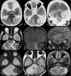
Tumour bleed manifesting as spontaneous extradural haematoma in posterior fossa
- PMID: 25008340
- PMCID: PMC4091297
- DOI: 10.1136/bcr-2014-205175
Tumour bleed manifesting as spontaneous extradural haematoma in posterior fossa
Abstract
We report a unique case of primary extradural angiosarcoma of posterior fossa manifesting as extradural haematoma in a 12-year-old boy who presented with acute onset headache, vomiting, nuchal rigidity and altered sensorium. The patient underwent a retromastoid suboccipital craniotomy on emergency basis, and the lesion was excised completely. Histopathology and immunohistochemistry revealed an angiosarcoma, following which radiation therapy was given. The patient showed complete clinical and neurological improvement. At a follow-up of 2 years he is in good health without any sign of regrowth.
2014 BMJ Publishing Group Ltd.
Figures


References
-
- Guode Z, Qi P, Hua G, et al. Primary cerebellopontine angle angiosarcoma. J Clin Neurosci 2008;15:942–46 - PubMed
-
- Kanai R, Kubota H, Terada T, et al. Spontaneous epidural hematoma due to skull metastasis of hepatocellular carcinoma. J Clin Neurosci 2009;16:137–40 - PubMed
-
- Hassan MF, Dhamija B, Palmer JD, et al. Spontaneous cranial extradural hematoma: case report and review of literature. Neuropathology 2009;29:480–4 - PubMed
Publication types
MeSH terms
LinkOut - more resources
Full Text Sources
Other Literature Sources
Medical
