. 2014 Oct 1;112(7):1739-47.
doi: 10.1152/jn.00383.2014. Epub 2014 Jul 9.

Hypersynchrony despite pathologically reduced beta oscillations in patients with Parkinson's disease: a pharmaco-magnetoencephalography study

Affiliations

Hypersynchrony despite pathologically reduced beta oscillations in patients with Parkinson's disease: a pharmaco-magnetoencephalography study

Elizabeth Heinrichs-Graham et al. J Neurophysiol. .

Abstract

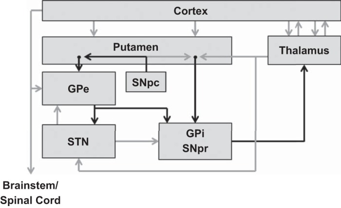
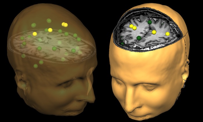
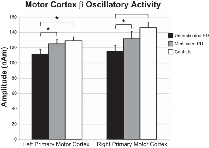
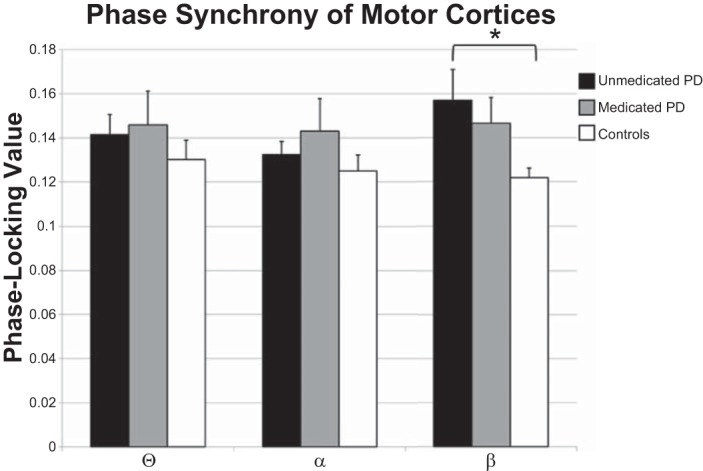
Parkinson's disease (PD) is a progressive debilitating neurodegenerative disorder clinically manifest by motor, posture and gait abnormalities. Human neurophysiological studies recording local field potentials within the subthalamic nucleus and scalp-based electroencephalography have shown pathological beta synchrony throughout the basal ganglia-thalamic-cortical motor network in PD. Notably, suppression of this pathological beta synchrony by dopamine replacement therapy or deep-brain stimulation has been associated with improved motor function. However, due to the invasive nature of these studies, it remains unknown whether this "pathological beta" is actually stronger than that observed in healthy demographically matched controls. We used magnetoencephalography to investigate neuronal synchrony and oscillatory amplitude in the beta range and lower frequencies during the resting state in patients with PD and a matched group of patients without neurological disease. Patients with PD were studied both in the practically defined drug "OFF" state, and after administration of dopamine replacements. We found that beta oscillatory amplitude was reduced bilaterally in the primary motor regions of unmedicated patients with PD compared with controls. Administration of dopaminergic medications significantly increased beta oscillatory activity, thus having a normalizing effect. Interestingly, we also found significantly stronger beta synchrony (i.e., hypersynchrony) between the primary motor regions in unmedicated patients with PD compared with controls, and that medication reduced this coupling which is in agreement with the intraoperative studies. These results are consistent with the known functionality of the basal ganglia-thalamic-cortical motor circuit and the likely consequences of beta hypersynchrony in the subthalamic nucleus of patients with PD.

Keywords: MEG; cortex; magnetoencephalography; oscillations; resting state.


Figures

Fig. 1.
Fig. 1.
Motor circuit components of importance to Parkinson's disease (PD). Black arrows indicate inhibitory [gamma-aminobutyric acid (GABA)-ergic] connections; gray arrows indicate excitatory (glutamatergic) connections. Cortex includes the cortical motor areas: primary motor, supplementary motor area (SMA), premotor areas, and cingulate motor areas. GPe, external segment of the globus pallidus; GPi, internal segment of the globus pallidus; SNpc, substantia nigra pars compacta; SNr, substantia nigra pars reticulata; STN, subthalamic nucleus. [Figure adapted from Delong and Wichmann (2007) with permission.]
Fig. 2.
Fig. 2.
Representation of the regional source model. For each participant, a 29-node (grid-point) model with dual orthogonal orientations was used to estimate regional neuronal activity during the resting-state magnetoencephalography recording using inverse spatial filtering. The model can be seen above overlaid on a structural MRI, with the yellow sources representing the cortical motor regions, which were of primary interest in the present study. The same three-dimensional rendition is shown in both the left and right panels, although the orientation of the image differs between the two panels to facilitate visualization of the spatial location of each source. The nonyellow color is only meant to aid in visually distinguishing the regional sources. Note that the regional sources are spaced equidistant apart, and that each represents activity over an extended cortical area (i.e., >1 cm3). Thus the time series of each node reflects the average neuronal activity over that brain region and not the amount of activation at a precise neuroanatomical coordinate (e.g., a voxel in Montreal Neurological Institute space). Following spectral analyses, the current amplitude (in nAm) from the dominant orientation of each source was used for statistical analysis.
Fig. 3.
Fig. 3.
Beta oscillatory amplitude differences in the primary motor regions. Amplitude (in nAm) is shown on the y-axis, while region is denoted along the x-axis. Significant differences between unmedicated patients with PD (black bars) and healthy controls (white bars) were restricted to the beta band in the left (P = 0.035) and right (P = 0.006) primary motor regions. Gray bars, data from medicated patients. Administration of anti-Parkinsonian medication increased the amplitude of beta activity in the left (P = 0.024) and right (P = 0.011) primary motor regions. A similar pattern of changes following medication was detected in the left and right SMA (not shown, see Table 2). Error bars indicate 1 SE. *P < 0.05.
Fig. 4.
Fig. 4.
Phase synchrony between the left and right primary motor regions. Phase-locking values are shown on the y-axis, while frequency band is denoted on the x-axis. Data for healthy controls are shown by white bars, for unmedicated patients with PD by black bars, and the data for medicated patients with PD by gray bars. Unmedicated patients with PD showed significantly stronger beta synchrony between the left and right primary motor regions than did healthy controls. While beta synchrony was reduced following medication, this reduction was not significant. *P < 0.05.

References

    1. Ashe J, Lungu OV, Basford AT, Lu X. Cortical control of motor sequences. Curr Opin Neurobiol 16: 213–221, 2006 - PubMed
    1. Baudrexel S, Witte T, Seifried C, von Wegner F, Beissner F, Klein JC, Steinmetz H, Deichmann R, Roeper J, Hilker R. Resting state fMRI reveals increased subthalamic nucleus-motor cortex connectivity in Parkinson's disease. Neuroimage 55: 1728–1738, 2011 - PubMed
    1. Becker KM, Heinrichs-Graham E, Fox HS, Robertson KR, Sandkovsky U, O'Neill J, Swindells S, Wilson TW. Decreased MEG beta oscillations in HIV-infected older adults during the resting state. J Neurovirol 19: 586–594, 2013 - PMC - PubMed
    1. Bosboom JL, Stoffers D, Stam CJ, van Dijk BW, Verbunt J, Berendse HW, Wolters E. Resting state oscillatory brain dynamics in Parkinson's disease: an MEG study. Clin Neurophysiol 117: 2521–2531, 2006 - PubMed
    1. Brookes MJ, Hale JR, Zumer JM, Stevenson CM, Francis ST, Barnes GR, Owen JP, Morris PG, Nagarajan SS. Measuring functional connectivity using MEG: methodology and comparison with fcMRI. Neuroimage 56: 1082–1104, 2011a - PMC - PubMed

Publication types