

Patterns of late gadolinium enhancement in Duchenne muscular dystrophy carriers
- PMID: 25008475
- PMCID: PMC4096415
- DOI: 10.1186/1532-429X-16-45
Patterns of late gadolinium enhancement in Duchenne muscular dystrophy carriers
Abstract
Background: This study was designed to assess whether cardiovascular magnetic resonance imaging (CMR) in Duchenne muscular dystrophy carriers (DMDc) may index any cell milieu elements of LV dysfunction and whether this cardiac phenotype may be related to genotype. The null hypothesis was that myocardial fibrosis, assessed by late gadolinium enhancement (LGE), might be similarly accounted for in DMDc and gender and age-matched controls.
Methods: Thirty DMDc patients had CMR and genotyping with 37 gender and age-matched controls. Systolic and diastolic LV function was assessed by 2D-echocardiography.
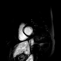
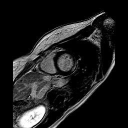
Results: Absolute and percent LGE were higher in muscular symptomatic (sym) than asymptomatic (asy) DMDc (1.77 ± 0.27 vs 0.76 ± 0.17 ml; F = 19.6, p < 0.0001 and 1.86 ± 0.26% vs 0.68 ± 0.17%, F = 22.1, p < 0.0001, respectively). There was no correlation between LGE and age. LGE was seen most frequently in segments 5 and 6; segment 5 was involved in all asy-DMDc. Subepicardial LGE predominated, compared to the mid-myocardial one (11 out of 14 DMDc). LGE was absent in the subendocardium. No correlations were seen between genotyping (type of mutation, gene region and protein domain), confined to the exon's study, and cardiac phenotype.
Conclusions: A typical myocardial LGE-pattern location (LV segments 5 and 6) was a common finding in DMDc. LGE was more frequently subepicardial plus midmyocardial in sym-DMDc, with normal LV systolic and diastolic function. No genotype-phenothype correlation was found.
Figures
Similar articles
-
Left ventricular systolic function and the pattern of late-gadolinium-enhancement independently and additively predict adverse cardiac events in muscular dystrophy patients.J Cardiovasc Magn Reson. 2014 Sep 25;16(1):81. doi: 10.1186/s12968-014-0081-1. J Cardiovasc Magn Reson. 2014. PMID: 25315351 Free PMC article.
-
Myocardial Fibrosis and Left Ventricular Dysfunction in Duchenne Muscular Dystrophy Carriers Using Cardiac Magnetic Resonance Imaging.Pediatr Cardiol. 2015 Oct;36(7):1495-501. doi: 10.1007/s00246-015-1192-7. Epub 2015 May 16. Pediatr Cardiol. 2015. PMID: 25976773
-
Cardiac involvement in female Duchenne and Becker muscular dystrophy carriers in comparison to their first-degree male relatives: a comparative cardiovascular magnetic resonance study.Eur Heart J Cardiovasc Imaging. 2016 Mar;17(3):326-33. doi: 10.1093/ehjci/jev161. Epub 2015 Jun 25. Eur Heart J Cardiovasc Imaging. 2016. PMID: 26113120
-
T1-Mapping and extracellular volume estimates in pediatric subjects with Duchenne muscular dystrophy and healthy controls at 3T.J Cardiovasc Magn Reson. 2020 Dec 10;22(1):85. doi: 10.1186/s12968-020-00687-z. J Cardiovasc Magn Reson. 2020. PMID: 33302967 Free PMC article.
-
Myocardial fibrosis imaging based on T1-mapping and extracellular volume fraction (ECV) measurement in muscular dystrophy patients: diagnostic value compared with conventional late gadolinium enhancement (LGE) imaging.Eur Heart J Cardiovasc Imaging. 2014 Sep;15(9):1004-12. doi: 10.1093/ehjci/jeu050. Epub 2014 Mar 30. Eur Heart J Cardiovasc Imaging. 2014. PMID: 24686257
Cited by
-
Cardiac Involvement in Women With Pathogenic Dystrophin Gene Variants.Front Neurol. 2021 Jul 27;12:707838. doi: 10.3389/fneur.2021.707838. eCollection 2021. Front Neurol. 2021. PMID: 34385974 Free PMC article.
-
Draft Guidance for Industry Duchenne Muscular Dystrophy, Becker Muscular Dystrophy, and Related Dystrophinopathies - Developing Potential Treatments for the Entire Spectrum of Disease.J Neuromuscul Dis. 2024;11(2):499-523. doi: 10.3233/JND-230219. J Neuromuscul Dis. 2024. PMID: 38363616 Free PMC article.
-
Identification of Cardiomyopathy-Associated Circulating miRNA Biomarkers in Muscular Dystrophy Female Carriers Using a Complementary Cardiac Imaging and Plasma Profiling Approach.Front Physiol. 2018 Dec 21;9:1770. doi: 10.3389/fphys.2018.01770. eCollection 2018. Front Physiol. 2018. PMID: 30622476 Free PMC article.
-
Duchenne Dilated Cardiomyopathy: Cardiac Management from Prevention to Advanced Cardiovascular Therapies.J Clin Med. 2020 Oct 1;9(10):3186. doi: 10.3390/jcm9103186. J Clin Med. 2020. PMID: 33019553 Free PMC article. Review.
-
Utilities and Limitations of Cardiac Magnetic Resonance Imaging in Dilated Cardiomyopathy.Korean J Radiol. 2023 Dec;24(12):1200-1220. doi: 10.3348/kjr.2023.0531. Korean J Radiol. 2023. PMID: 38016680 Free PMC article. Review.
References
-
- Martinez HR, Pignatelli R, Belmont JW, Craigen WJ, Jefferies JL. Childhood onset of left ventricular dysfunction in a female manifesting carrier of muscular dystrophy. Am J Med Genet. 2011;155:3025–3029. - PubMed
-
- Nolan MA, Jones ODH, Pedersen RL, Johnston HM. Cardiac assessment in childhood carriers of Duchenne and Becker muscular dystrophies. Neuromusc Disord. 2003;13:129–132. - PubMed
-
- Mirabella M, Servidei S, Manfredi G, Ricci E, Frustaci A, Bertini E, Rana M, Tonali P. Cardiomyopathy may be the only clinical manifestation in female carriers of Duchenne muscular dystrophy. Neurology. 1993;43:2342–2345. - PubMed
-
- Kamakura K, Kawai M, Arahata K, Koizumi H, Watanabe K, Sugita H. A manifesting carrier of Duchenne muscular dystrophy with severe myocardial symptoms. J Neurol. 1990;237:483–485. - PubMed
-
- Hoogerwaard EM, van der Wouw PA, Wilde AA, Bakker E, Ippel PF, Oosterwijk JC, Majoor-Krakauer DF, van Essen AJ, Leschot NJ, de Visser M. Cardiac involvement in carriers of Duchenne and Becker muscular dystrophy. Neuromuscul Disord. 1999;9:347–351. - PubMed
MeSH terms
Substances
LinkOut - more resources
Full Text Sources
Other Literature Sources
