. 2015;24(8):1571-84.
doi: 10.3727/096368914X682792. Epub 2014 Jul 8.

Diabetic Mesenchymal Stem Cells Are Ineffective for Improving Limb Ischemia Due to Their Impaired Angiogenic Capability

Affiliations

Diabetic Mesenchymal Stem Cells Are Ineffective for Improving Limb Ischemia Due to Their Impaired Angiogenic Capability

Hyongbum Kim et al. Cell Transplant. 2015.

Abstract

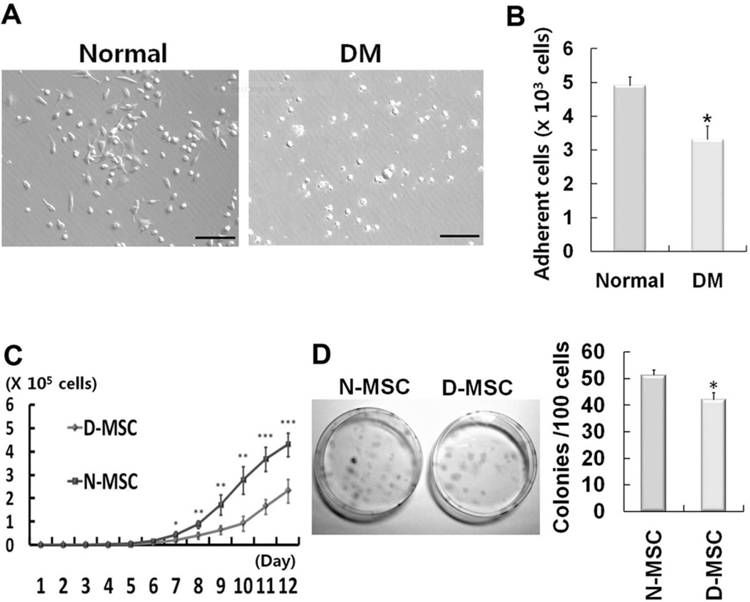
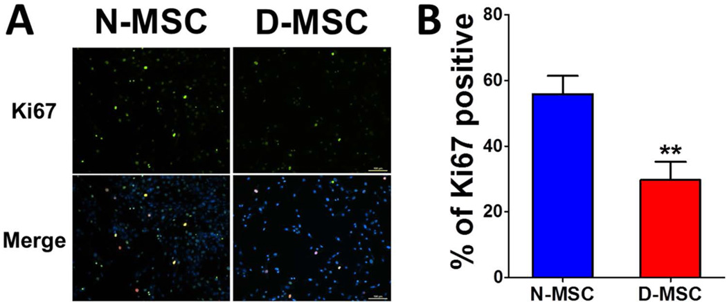
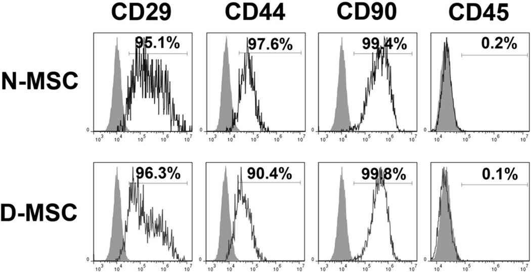
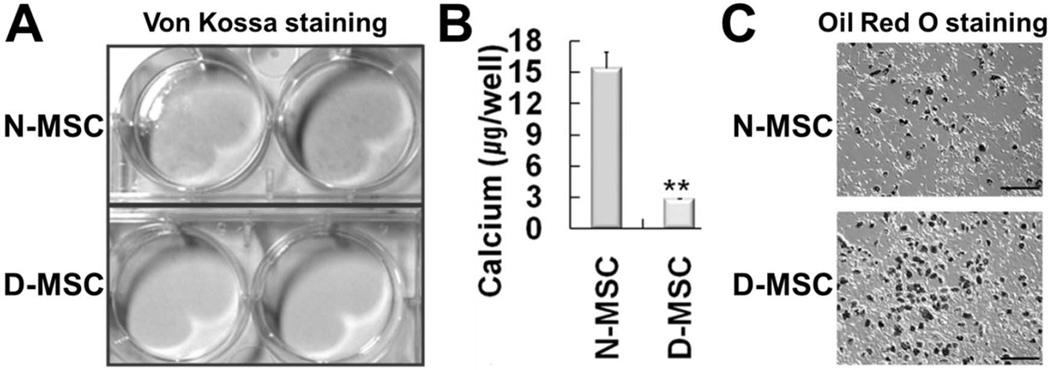
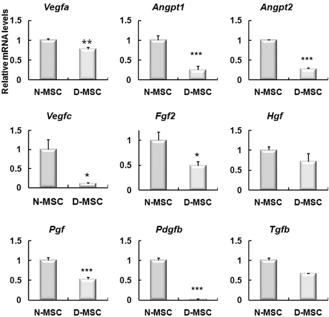
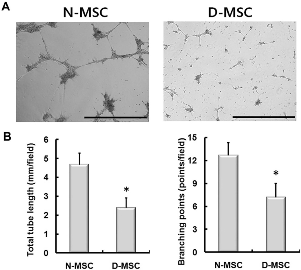
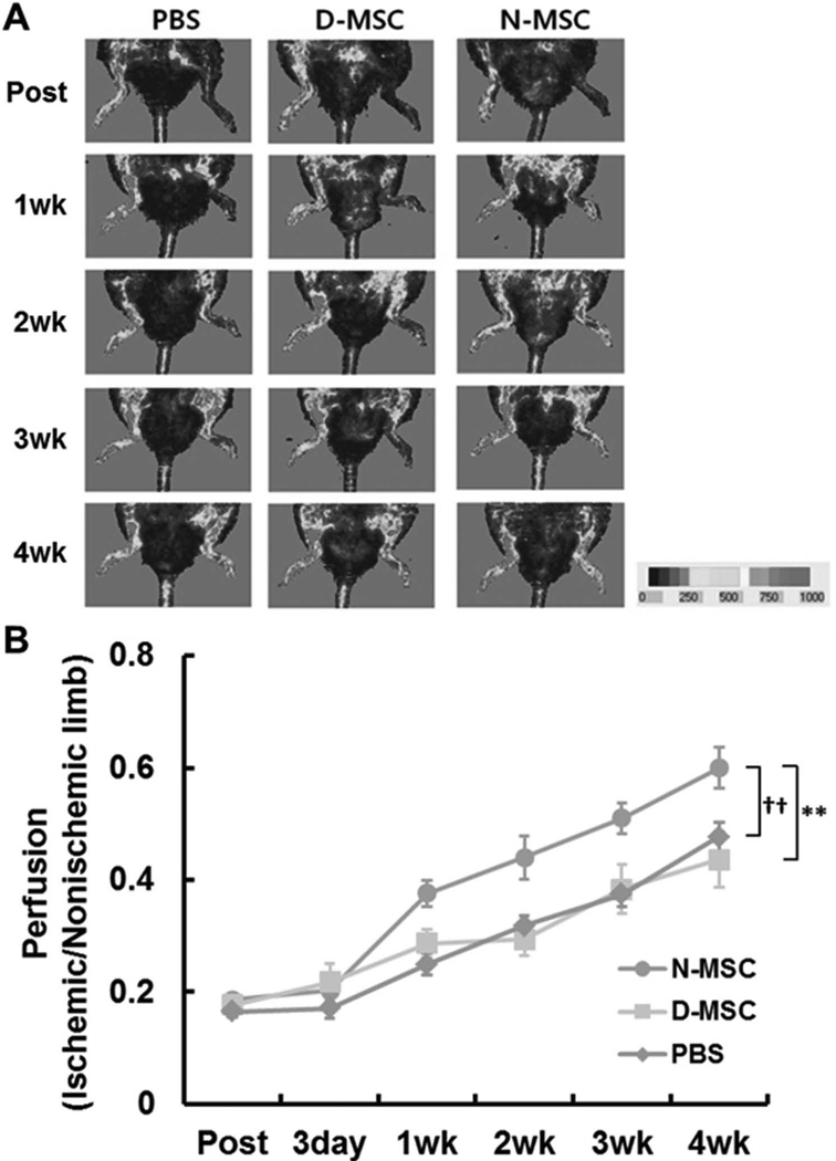
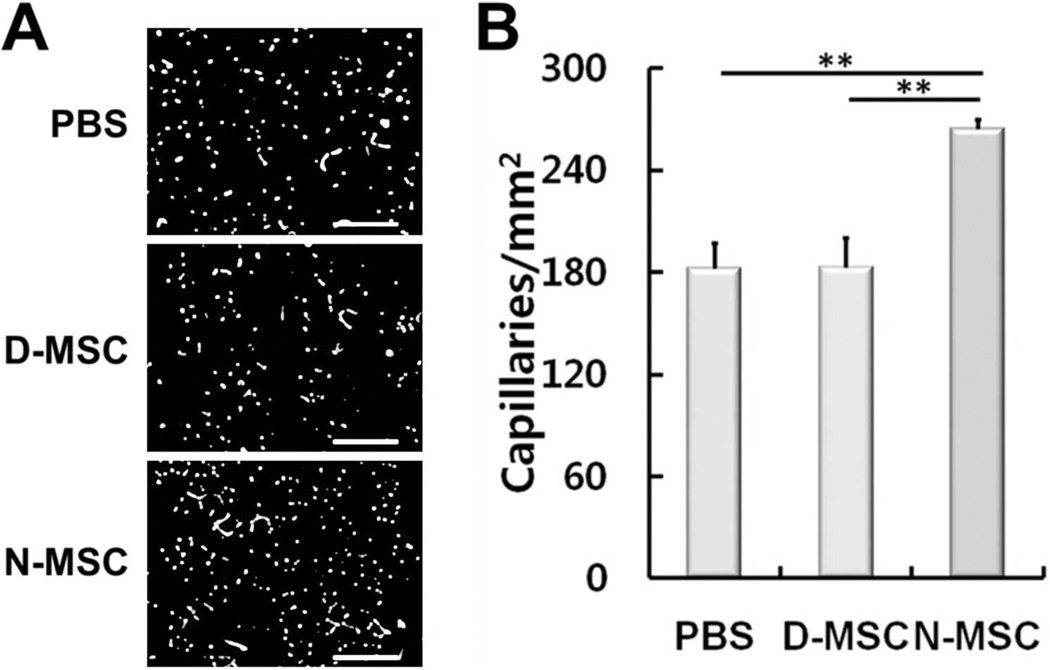
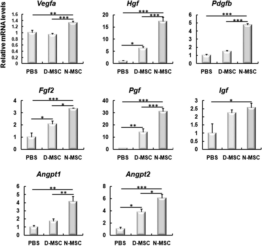
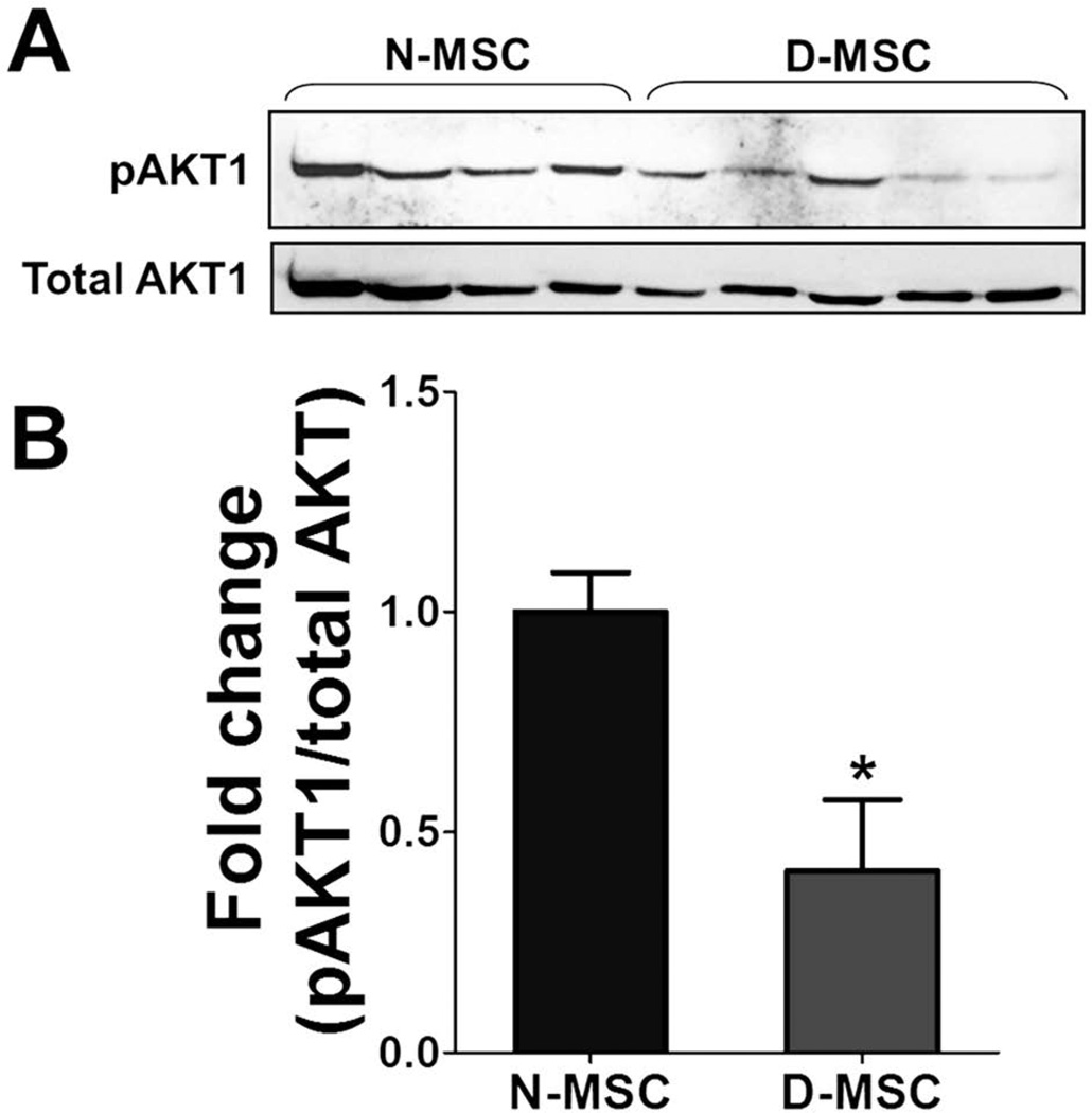
The purpose of this study was to investigate the effects of diabetes on mesenchymal stem cells (MSCs) in terms of their angiogenic and therapeutic potential for repairing tissue ischemia. We culture-isolated MSCs from streptozotocin-induced diabetic rats (D-MSCs) and compared their proliferation, differentiation, and angiogenic effects with those from normal rats (N-MSCs). The angiogenic effects of MSCs were evaluated by real-time PCR, in vitro tube formation assay, and transplantation of the MSCs into a hindlimb ischemia model followed by laser Doppler perfusion imaging. The number of MSCs derived from diabetic rats was smaller, and their proliferation rate was slower than N-MSCs. Upon induction of differentiation, the osteogenic and angiogenic differentiation of D-MSCs were aberrant compared to N-MSCs. The expression of angiogenic factors was lower in D-MSCs than N-MSCs. D-MSCs cocultured with endothelial cells resulted in decreased tube formation compared to N-MSCs. D-MSCs were ineffective to improve hindlimb ischemia and showed lower capillary density and angiogenic gene expression in ischemic limbs than N-MSCs. D-MSCs have defective proliferation and angiogenic activities and are ineffective for repairing hindlimb ischemia. Newer measures are needed before MSCs can be employed as a source for autologous cell therapy.


Conflict of interest statement

The authors declare no conflicts of interest.

Figures

Figure 1
Figure 1
Impaired proliferation of mesenchymal stem cells (MSCs) derived from diabetic rats. (A) Representative photomicrographs of MSC culture at 4 days. Most adherent cells from normal rats were spindle shaped, whereas many of those from diabetic rats (DM) were rounded up, suggestive of aberrancy. Scale bars: 100 µm. (B) The numbers of adherent cells, most of which are MSCs, were significantly higher in the normal rat group than the DM group (*p < 0.05, n = 3) at 10 days of culture. (C) Growth curve of MSCs. The numbers of MSCs were counted daily. D-MSCs showed impaired proliferation compared to N-MSCs. (*p < 0.05, **p < 0.01, ***p < 0.001, n = 4). (D) One hundred MSCs were plated per 100-mm dish, and the number of colonies was counted at 14 days using crystal violet staining. The number of colony-forming cells was significantly lower in D-MSCs than N-MSCs (*p < 0.05, n = 4).
Figure 2
Figure 2
Decreased Ki-67-positive cells in D-MSCs. Culture-expanded N-MSCs and D-MSCs were immunostained with anti-Ki-67 antibody to examine their proliferation capacity. (A) Representative immunocytochemical findings for Ki-67-positive cells. The nuclei of the proliferating cells were demonstrated by double positivity for green (Ki-67) and blue fluorescence (DAPI). Scale bar: 100 µm. (B) Percentages of Ki-67-positive cells in N-MSC and D-MSC (**p < 0.01).
Figure 3
Figure 3
Flow cytometric characterization of N- and D-MSCs. MSCs were culture isolated from BM of normal and diabetic rats. N-MSCs and D-MSCs were equally positive for MSC markers such as CD29, CD44, and CD90 and were negative for a pan-hematopoietic cell marker, CD45. Gray curves indicate the isotype controls.
Figure 4
Figure 4
D-MSCs have aberrant differentiation propensity. von Kossa staining (A) and quantification of the deposited calcium (B) after osteogenic differentiation of N- or D-MSCs. D-MSCs showed weaker von Kossa staining and lower levels of deposited calcium than N-MSCs (**p < 0.01, n = 3), suggesting impaired osteogenic differentiation. (C) Oil red O staining after adipogenic differentiation for 3 weeks. D-MSCs showed stronger Oil red O staining than N-MSCs, suggesting enhanced adipogenic differentiation. Scale bars: 400 µm.
Figure 5
Figure 5
The expression levels of angiogenic genes were lower in D-MSCs than N-MSCs. Real-time RT-PCR showed that the expression levels of Vegfa, Angpt1, Angpt2, Vegfc, Fgf2, Pgf, and Pdgfb were significantly lower in D-MSCs than N-MSCs (*p < 0.05, **p < 0.01, ***p < 0.001), whereas the levels of Hgf and Tgfb were not significantly different between the two groups. n = 4–6.
Figure 6
Figure 6
In vitro tube formation by coculture of MSCs and endothelial cells. Human umbilical vein endothelial cells, 7 × 10⁴, were cocultured with 2.5 × 10⁵ N- or D-MSCs and plated onto Matrigel™. Tube formation was observed after 16 h. (A) Representative photomicrographs showing the in vitro tube formation. (B) The total tube length and number of branching points of the tubular network per field were higher in the N-MSC group than the D-MSC group (*p < 0.05, n = 6). Scale bars: 1 mm.
Figure 7
Figure 7
Impaired improvement of hindlimb ischemia by D-MSCs. Three million N- or D-MSCs were intramuscularly injected into the ischemic hindlimbs. Blood perfusion of the limb was determined using LDPI. Representative LDPI images (A) and quantification of the perfusion (B) showed that hindlimb blood flow was not significantly enhanced in the D-MSC group compared to the PBS group. The N-MSC group showed higher blood flow compared to the D-MSC group (**p < 0.01) and the PBS group (††p < 0.01). n = 5–14.
Figure 8
Figure 8
Impaired neovascularization in hindlimb ischemia by D-MSCs. Four weeks after implantation of N- or D-MSCs into ischemic limbs, the limb tissues were harvested and stained with ILB4. Representative fluorescent micrographs (A) and quantification of the capillary density (B) showed that capillary density was not increased in the D-MSCs compared to the PBS control, but did increase in the N-MSC group compared to the D-MSC (**p < 0.01) and PBS groups (**p < 0.01). n = 4. Scale bars: 200 µm.
Figure 9
Figure 9
Upregulation of angiogenic, antiapoptotic, and vessel-stabilizing factors in ischemic hindlimbs injected with D-MSCs and controls. The hindlimb tissues were harvested 2 weeks after cell implantation. Total RNA was isolated and subjected to quantitative RT-PCR. Gene levels were normalized to GAPDH. Data are presented as fold differences compared to the value of the PBS group. *p < 0.05, **p < 0.01, ***p < 0.001, n = 3–8.
Figure 10
Figure 10
ATK1 levels in N- and D-MSCs. (A) Total proteins were isolated from N-MSCs derived from four normal rats and D-MSCs from five diabetic rats and subjected to Western blotting analysis. (B) The levels of phosphorylated AKT1 normalized to total AKT1 were lower in D-MSCs than in N-MSCs (*p < 0.05; n = 4 for N-MSCs, n = 5 for D-MSCs).

References

    1. Ackah E, Yu J, Zoellner S, Iwakiri Y, Skurk C, Shibata R, Ouchi N, Easton RM, Galasso G, Birnbaum MJ, Walsh K, Sessa WC. Akt1/protein kinase Balpha is critical for ischemic and VEGF-mediated angiogenesis. J. Clin. Invest. 2005;115(8):2119–2127. - PMC - PubMed
    1. Al-Khaldi A, Eliopoulos N, Martineau D, Lejeune L, Lachapelle K, Galipeau J. Postnatal bone marrow stromal cells elicit a potent VEGF-dependent neoangiogenic response in vivo. Gene Ther. 2003;10(8):621–629. - PubMed
    1. Amado LC, Saliaris AP, Schuleri KH, St John M, Xie JS, Cattaneo S, Durand DJ, Fitton T, Kuang JQ, Stewart G, Lehrke S, Baumgartner WW, Martin BJ, Heldman AW, Hare JM. Cardiac repair with intramyocardial injection of allogeneic mesenchymal stem cells after myocardial infarction. Proc. Natl. Acad. Sci. USA. 2005;102(32):11474–11479. - PMC - PubMed
    1. Baddoo M, Hill K, Wilkinson R, Gaupp D, Hughes C, Kopen GC, Phinney DG. Characterization of mesenchymal stem cells isolated from murine bone marrow by negative selection. J. Cell. Biochem. 2003;89(6):1235–1249. - PubMed
    1. Brasacchio D, Okabe J, Tikellis C, Balcerczyk A, George P, Baker EK, Calkin AC, Brownlee M, Cooper ME, El-Osta A. Hyperglycemia induces a dynamic cooperativity of histone methylase and demethylase enzymes associated with gene-activating epigenetic marks that coexist on the lysine tail. Diabetes. 2009;58(5):1229–1236. - PMC - PubMed

Publication types

MeSH terms

Substances

LinkOut - more resources