. 2014 Sep;88(18):10909-17.
doi: 10.1128/JVI.01910-14. Epub 2014 Jul 9.

Type III interferon attenuates a vesicular stomatitis virus-based vaccine vector

Affiliations

Type III interferon attenuates a vesicular stomatitis virus-based vaccine vector

Ryann C Guayasamin et al. J Virol. 2014 Sep.

Abstract

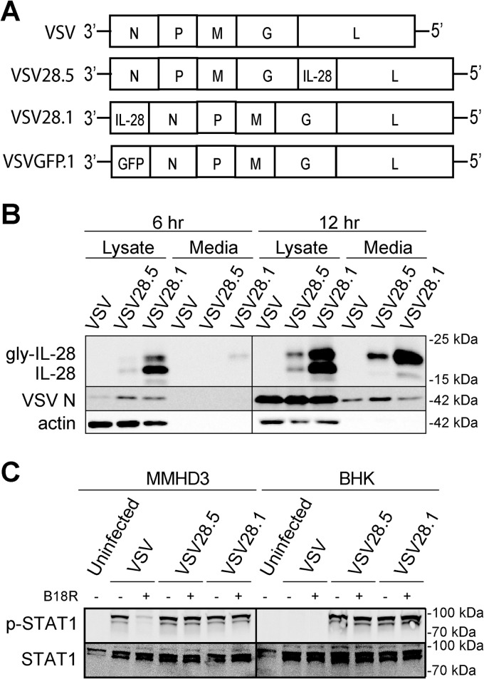
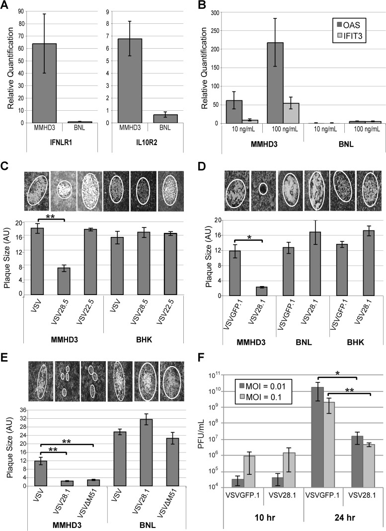
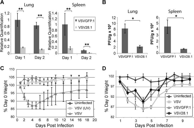
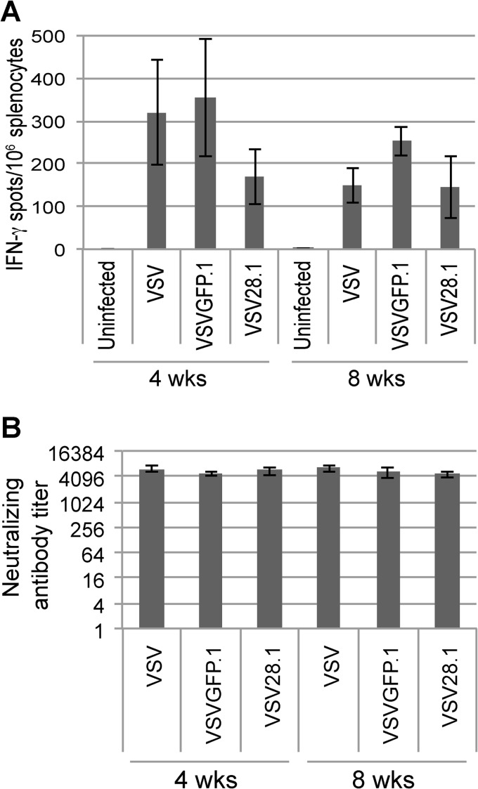
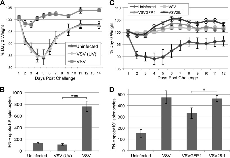
Vesicular stomatitis virus (VSV) has been extensively studied as a vaccine vector and oncolytic agent. Nevertheless, safety concerns have limited its widespread use in humans. The type III lambda interferon (IFN-λ) family of cytokines shares common signaling pathways with the IFN-α/β family and thus evokes similar antiviral activities. However, IFN-λ signals through a distinct receptor complex that is expressed in a cell type-specific manner, which restricts its activity to epithelial barriers, particularly those corresponding to the respiratory and gastrointestinal tracts. In this study, we determined how IFN-λ expression from recombinant VSV would influence vector replication, spread, and immunogenicity. We demonstrate that IFN-λ expression severely attenuates VSV in cell culture. In vivo, IFN-λ limits VSV replication in the mouse lung after intranasal administration and reduces virus spread to other organs. Despite this attenuation, however, the vector retains its capacity to induce protective CD8 T cell and antibody responses after a single immunization. These findings demonstrate a novel method of viral vector attenuation that could be used in both vaccine and oncolytic virus applications.

Importance: Viruses such as VSV that are used as vaccine vectors can induce protective T cell and antibody responses after a single dose. Additionally, IFN-λ is a potent antiviral agent that has certain advantages for clinical use compared to IFN-α/β, such as fewer patient side effects. Here, we demonstrate that IFN-λ attenuates VSV replication and spread following intranasal virus delivery but does not reduce the ability of VSV to induce potent protective immune responses. These findings demonstrate that the type III IFN family may have widespread applicability for improving the safety and efficacy of viral vaccine and oncolytic vectors.


Figures

FIG 1
FIG 1
Expression of functionally active IFN-λ (IL-28) protein from VSV. (A) Genomic diagrams of the VSV vectors generated in this study. All five genes encoding the nucleocapsid (N), the phosphoprotein (P), the matrix protein (M), the glycoprotein (G), and the RNA-dependent RNA polymerase (L) were present in the viruses. The mouse IFN-λ2 (IL-28A) gene was inserted either upstream of the L gene in the 5th position (VSV28.5) or upstream of the N gene in the 1st position (VSV28.1). Control viruses with either no insert (VSV) or the GFP gene inserted in the 1st position (VSVGFP.1) are also depicted. (B) Immunoblot analysis of glycosolated or unglycosolated IFN-λ protein expressed in the lysates or media collected from BHK cells infected with the designated viruses at either 6 or 12 h p.i. Molecular mass values are indicated. (C) Immunoblot of STAT1 (p-STAT1α [91 kDa] and a splice variant, STAT1β [84 kDa]) phosphorylation in MMHD3 cells incubated for 30 min with media from either MMHD3 or BHK cells collected after a 24-h infection with VSV, VSV28.5, or VSV28.1. Media were treated with or without soluble B18R protein to block IFN-α/β before transfer onto MMHD3 cells as designated. Molecular mass values are indicated.
FIG 2
FIG 2
VSV expressing IFN-λ is attenuated in IFN-λ-responsive cells. (A) Total RNA was prepared from MMHD3 or BNL cells, and IL-10Rβ and IFN-λR1 mRNA was quantified by quantitative RT-PCR. (B) MMHD3 or BNL cells were treated with or without 10 ng/ml or 100 ng/ml IFN-λ for 24 h and harvested for total RNA isolation. ISGs OAS and IFIT3 were quantified by quantitative RT-PCR. (C to E) MMHD3, BNL, or BHK cells were infected with serially diluted virus, and at 2 days p.i., plaques were stained and then visualized and photographed under a light microscope for quantification of the average relative plaque size by measuring the area of an ellipse that was fitted to the plaque. The bar graphs depict the mean individual plaque areas, with error bars representing the standard errors of the means. Representative images of plaques are shown above each graph. (F) MMHD3 cells were infected with VSVGFP.1 or VSV28.1 at MOIs of 0.01 and 0.1, and media were collected at 10 and 24 h p.i. to quantify viral production by a standard plaque assay. *, P < 0.05; **, P < 0.01.
FIG 3
FIG 3
Reduced replication and spread of VSV28.1 following intranasal infection of mice. (A and B) Mice were intranasally infected with 1 × 10⁶ PFU of VSVGFP.1 or VSV28.1. The lungs and spleen were harvested, and viral genomic RNA (VSV-N) (A) or viral titers (B) were quantified by quantitative RT-PCR or by a plaque assay, respectively. (C and D) Mice were left uninfected or intranasally infected with 1 × 10⁶ PFU of VSV or its UV-inactivated equivalent (C) or 1 × 10⁶ PFU of VSV, VSVGFP.1, or VSV28.1 (D). Average weight loss was measured daily until mice had recovered. Error bars represent the standard errors of the means (n = 5). *, P < 0.05; **, P < 0.01.
FIG 4
FIG 4
VSV28.1 induces CD8 T cell and antibody responses similar to those of nonattenuated vectors. Mice were intranasally infected with 1 × 10⁶ PFU of VSV, VSVGFP.1, or VSV28.1. At 4 and 8 weeks p.i., the splenic VSV-specific CD8 T cells were quantified by IFN-γ ELISPOT assays (A), and VSV neutralizing antibody titers were measured in the serum (B). Error bars represent standard errors of the means (n = 5).
FIG 5
FIG 5
VSV28.1 immunization protects from subsequent virus challenge. (A and C) Mice were immunized intranasally with PBS (Uninfected), 1 × 10⁶ PFU of VSV, or the equivalent of 1 × 10⁶ PFU of UV-inactivated VSV (A) or with PBS, VSV, VSVGFP.1, or VSV28.1 (C). At 1 month p.i., all groups were challenged with 1 × 10⁶ PFU of VSV(G)ch, and weight loss was measured for 2 weeks postchallenge. The average weight for each group is shown. (B and D) Splenocytes collected from mice 2 weeks postchallenge were assayed to quantify splenic VSV-specific CD8 T cells by IFN-γ ELISPOT assays (n = 5). *, P < 0.05; ***, P < 0.001.

Similar articles

Cited by

References

    1. Finke S, Conzelmann KK. 2005. Recombinant rhabdoviruses: vectors for vaccine development and gene therapy. Curr. Top. Microbiol. Immunol. 292:165–200. 10.1007/3-540-27485-5_8 - DOI - PubMed
    1. Lawson ND, Stillman EA, Whitt MA, Rose JK. 1995. Recombinant vesicular stomatitis viruses from DNA. Proc. Natl. Acad. Sci. U. S. A. 92:4477–4481. 10.1073/pnas.92.10.4477 - DOI - PMC - PubMed
    1. Schnell MJ, Buonocore L, Kretzschmar E, Johnson E, Rose JK. 1996. Foreign glycoproteins expressed from recombinant vesicular stomatitis viruses are incorporated efficiently into virus particles. Proc. Natl. Acad. Sci. U. S. A. 93:11359–11365. 10.1073/pnas.93.21.11359 - DOI - PMC - PubMed
    1. Schnell MJ, Buonocore L, Whitt MA, Rose JK. 1996. The minimal conserved transcription stop-start signal promotes stable expression of a foreign gene in vesicular stomatitis virus. J. Virol. 70:2318–2323 - PMC - PubMed
    1. Tesh RB, Peralta PH, Johnson KM. 1969. Ecologic studies of vesicular stomatitis virus. I. Prevalence of infection among animals and humans living in an area of endemic VSV activity. Am. J. Epidemiol. 90:255–261 - PubMed

Publication types

MeSH terms

LinkOut - more resources