. 2014 Sep:54:49-59.
doi: 10.1016/j.biocel.2014.06.013. Epub 2014 Jul 4.

Androgen receptor and its splice variant, AR-V7, differentially regulate FOXA1 sensitive genes in LNCaP prostate cancer cells

Affiliations

Androgen receptor and its splice variant, AR-V7, differentially regulate FOXA1 sensitive genes in LNCaP prostate cancer cells

William C Krause et al. Int J Biochem Cell Biol. 2014 Sep.

Abstract

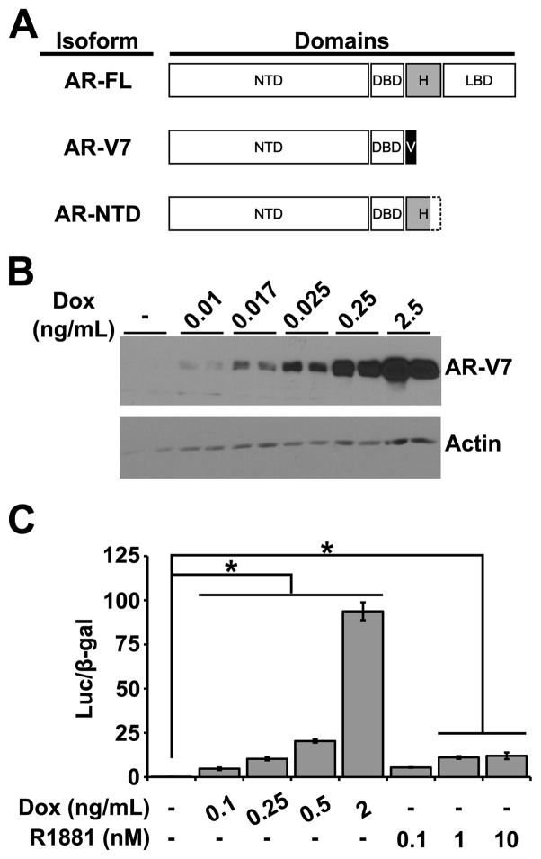
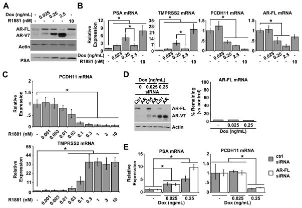
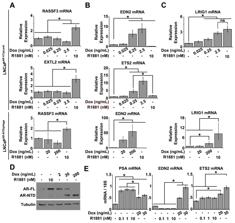
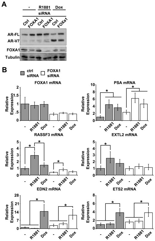
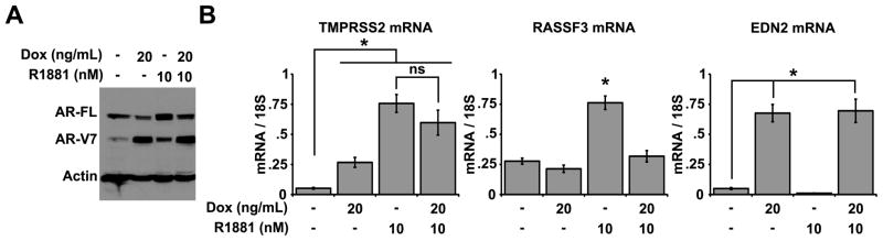
Prostate cancer (PCa) is an androgen-dependent disease, and tumors that are resistant to androgen ablation therapy often remain androgen receptor (AR) dependent. Among the contributors to castration-resistant PCa are AR splice variants that lack the ligand-binding domain (LBD). Instead, they have small amounts of unique sequence derived from cryptic exons or from out of frame translation. The AR-V7 (or AR3) variant is constitutively active and is expressed under conditions consistent with CRPC. AR-V7 is reported to regulate a transcriptional program that is similar but not identical to that of AR. However, it is unknown whether these differences are due to the unique sequence in AR-V7, or simply to loss of the LBD. To examine transcriptional regulation by AR-V7, we have used lentiviruses encoding AR-V7 (amino acids 1-627 of AR with the 16 amino acids unique to the variant) to prepare a derivative of the androgen-dependent LNCaP cells with inducible expression of AR-V7. An additional cell line was generated with regulated expression of AR-NTD (amino acids 1-660 of AR); this mutant lacks the LBD but does not have the AR-V7 specific sequence. We find that AR and AR-V7 have distinct activities on target genes that are co-regulated by FOXA1. Transcripts regulated by AR-V7 were similarly regulated by AR-NTD, indicating that loss of the LBD is sufficient for the observed differences. Differential regulation of target genes correlates with preferential recruitment of AR or AR-V7 to specific cis-regulatory DNA sequences providing an explanation for some of the observed differences in target gene regulation.

Keywords: Androgen receptor; Cell biology; Nuclear receptors; Prostate cancer; Transcription.


Figures

FIGURE 1
FIGURE 1. Inducible expression of constitutively active AR variants
A, schematic illustration of the differences in functional domains among AR-FL, AR-V7, and AR-NTD. N-terminal domain (NTD), DNA binding domain (DBD), hinge (H), variant sequence (V), and ligand binding domain (LBD). B, LNCaPAR-V7/pLenti cells were treated in duplicate with indicated concentrations of Dox for 24 hours. Western blots were probed with antibodies against AR-V7 (upper) or Actin (lower). C, cells were treated as indicated for 24 hours. Transcriptional activities of AR-FL and AR-V7 were measured using a transiently transfected AR responsive Luciferase reporter and normalized to β-gal activity. Error bars indicate SEM. n = 3. Statistical differences between groups were determined by ANOVA with Bonferroni post-hoc test. *, p < 0.01.
FIGURE 2
FIGURE 2. Analysis of AR-V7 regulation of endogenous targets
A, inducible LNCaPAR-V7/pLenti cells were treated for 24 hours with the indicated amounts of vehicle control, Dox, or R1881. Changes in protein level were analyzed by Western blot using antibodies that recognize: total AR (upper), Actin (middle), or PSA (lower). B, cells were treated in parallel with those in A and changes in expression of the indicated genes were quantified by qPCR. C, parental LNCaP cells were treated with increasing concentrations of R1881 and changes in expression of the indicated target genes were quantified by qPCR. DE, LNCaPAR-V7/pLenti cells in complete medium were transiently transfected with siRNA targeting AR-FL. Medium was replaced with charcoal-stripped serum and cells were treated as indicated. Depletion of AR-FL protein (D left) and transcript (D right) was monitored by Western blot using a pan-AR antibody and by qPCR, respectively. In parallel, changes in target gene expression following induction of AR-V7 were quantified by qPCR (E). For all panels, error bars indicate the upper and lower bounds of the 95% confidence interval. n ≥ 3 experiments performed in triplicate. Statistical differences between groups were determined by ANOVA with Bonferroni post-hoc test (B and E), or between treatment and control groups by Dunnett’s multiple comparison test (C). *, p < 0.01.
FIGURE 3
FIGURE 3. Differential regulation by AR and its variants of target genes that are co-regulated by FOXA1
AC, LNCaPAR-V7/pLenti or LNCaPAR-NTD/pHage cells were treated, as indicated, for 24 hours and transcripts measured by qPCR. The activities of AR-FL, AR-V7, and AR-NTD were compared across a subset of target genes that have been previously demonstrated to require (A and C) or be inhibited (B) by the expression of FOXA1. Error bars indicate the upper and lower bounds of the 95% confidence interval. n ≥ 3 experiments performed in triplicate. D, expression levels of AR-NTD in the LNCaPAR-NTD/pHage line were determined by Western blot using the pan-AR antibody. E, LNCaPAR-V7/pHage cells were treated for 24 hours, and expression of transcripts measured by qPCR. Error bars indicate standard error of the mean. n ≥ 3 experiments performed in triplicate. For all qPCR data, statistical differences between groups were determined by ANOVA with Bonferroni post-hoc test. *, p < 0.01.
FIGURE 4
FIGURE 4. Selective Regulation of target genes by AR-FL, but not AR-V7, depends on FOXA1
48 hours after transfection with siRNA targeting FOXA1 or a nonspecific control sequence LNCaPAR-V7/pHage cells were placed in 10% charcoal-stripped serum, and treated for an additional 24 hours with 10 nM R1881 or 20 ng/mL Dox, as indicated. A, Protein levels of AR-FL, AR-V7, and FOXA1 were assessed by Western blot using the pan-AR and FOXA1 antibodies. B, expression of the indicated target genes was measured by qPCR. Error bars indicate the upper and lower bounds of the 95% confidence interval. n = 3 experiments performed in triplicate. Statistical differences within groups transfected with the same siRNA were determined by one-way ANOVA with Bonferroni post-hoc test. *, p < 0.01.
FIGURE 5
FIGURE 5. Variant-specific target genes are regulated in multiple cell lines with inducible or endogenous expression of AR-V7
A–B, VCaPAR-V7/pHage cells in 10% charcoal-stripped serum were treated for 24 hours with the indicated amounts of Dox or R1881. AR-FL and AR-V7 protein levels were assessed by Western blot using the AR-441 antibody (A), and in parallel the relative expression levels of the given target genes were determined by qPCR (B). Error bars indicate the upper and lower bounds of the 95% confidence interval. C, 22Rv-1, C4-2, and LNCaP PCa cell lines were maintained in charcoal stripped-serum for 48 hours and then treated with vehicle or 10 nM R1881 for 24 hours. Endogenous expression levels of AR-FL and AR-V7 were compared by Western blot using the AR-441 antibody. D, LNCaP, C4-2, and 22Rv-1 cells were maintained in charcoal-stripped serum for 48 hours and expression of the indicated target genes was measured by qPCR. Error bars indicate the upper and lower bounds of the 95% confidence interval. n = 3 experiments performed in triplicate. Statistical differences between groups were determined by ANOVA with Bonferroni post-hoc test. *, p < 0.01. E–F, 22Rv-1 cells in complete medium were transiently transfected with pan-AR or control siRNA for 48 hours. AR protein levels were measured by Western blot using the pan-AR antibody (E) and changes in expression of the indicated target genes were analyzed by qPCR (F). Error bars indicate standard error of the mean. n = 3 experiments performed in triplicate. Statistical differences between groups were determined by one-way Student’s T-test. *, p < 0.01.
FIGURE 6
FIGURE 6. AR-FL and AR-V7 preferentially recognize distinct cis-regulatory binding sites
LNCaPAR-V7/pLenti were treated for 24 hours with 0.25 ng/mL Dox or 10 nM R1881. AR recruitment to the indicated sites was measured by ChIP-qPCR using the AR-N20 antibody, which recognizes all AR forms (grey bars), the AR-V7 antibody (black bars) or a control IgG antibody (open bars). Enrichment of the binding site sequence in response to either treatment was quantified as a percentage of the input amount. A, ChIP-qPCR analysis of AR-FL and AR-V7 binding to the PSA enhancer. BC, upper panels show UCSC Genome browser view of the genomic regions surrounding RASSF3 (B) and EDN2 (C). Vertical bars in overlaid tracts indicate peaks of binding events by FOXA1 and AR, both in the presence or absence of FOXA1, as captured by Wang et al. (2011) (GSE27682). Labeled binding sites (12-529, 12-89, and 1-223) were analyzed for AR recruitment in response to isoform-specific treatments by ChIP-qPCR (lower panels). Error bars indicate the SEM. n ≥ 3 experiments. Statistical differences between control and activating treatments when pulled-down with the AR-N20 antibody were determined by ANOVA with Bonferroni post hoc test. *, p < 0.05. The effect of treatment in the AR-V7 ChIP was determined by two-way Student’s T-test. *, p < 0.05. D, Comparison of the overlap between AR and FOXA1 cis-regulatory elements on target genes that are specifically activated by AR-V7 (ETS2) or by AR-FL (EXTL2).
FIGURE 7
FIGURE 7. AR-V7 antagonizes the activity of AR-FL on a subset of genes with distinct receptor binding sites
LNCaPAR-V7/pHage cells in 10% charcoal-stripped serum were treated for 24 hours with Dox or R1881. In the combined treatment groups Dox was added 6 hours prior to R1881, to allow time for expression of AR-V7. A, total AR protein levels in response to treatment were assessed by Western blot using a pan-AR antibody; blots were also probed for Actin expression to ensure equal loading. B, mRNA expression of the indicated target genes was quantified by qPCR. Error bars indicate the SEM. n ≥ 3 experiments performed in triplicate. Statistical differences between groups were determined by ANOVA with Bonferroni post-hoc test. *, p < 0.01.

Similar articles

Cited by

References

    1. Agoulnik IU, Bingman WE, Nakka M, Li W, Wang Q, Liu XS, et al. Target gene-specific regulation of androgen receptor activity by p42/p44 mitogen-activated protein kinase. Mol Endocrinol. 2008 Nov;22(11):2420–32. - PMC - PubMed
    1. Agoulnik IU, Krause WC, Bingman WE, Rahman HT, Amrikachi M, Ayala GE, et al. Repressors of androgen and progesterone receptor action. J Biol Chem. 2003 Aug 15;278(33):31136–48. - PubMed
    1. Agoulnik IU, Vaid A, Bingman WE, Erdeme H, Frolov A, Smith CL, et al. Role of SRC-1 in the promotion of prostate cancer cell growth and tumor progression. Cancer Res. 2005 Sep 1;65(17):7959–67. - PubMed
    1. Agoulnik IU, Vaid A, Nakka M, Alvarado M, Bingman WE, Erdem H, et al. Androgens modulate expression of transcription intermediary factor 2, an androgen receptor coactivator whose expression level correlates with early biochemical recurrence in prostate cancer. Cancer Res. 2006 Nov 1;66(21):10594–602. - PubMed
    1. Bai W, Weigel NL. Phosphorylation of Ser211 in the chicken progesterone receptor modulates its transcriptional activity. J Biol Chem. 1996 May 31;271(22):12801–6. - PubMed

Publication types

MeSH terms