. 2014 Jul 9;6(244):244re5.
doi: 10.1126/scitranslmed.3008882.

Plasmodium falciparum transmission stages accumulate in the human bone marrow

Affiliations

Plasmodium falciparum transmission stages accumulate in the human bone marrow

Regina Joice et al. Sci Transl Med. .

Abstract

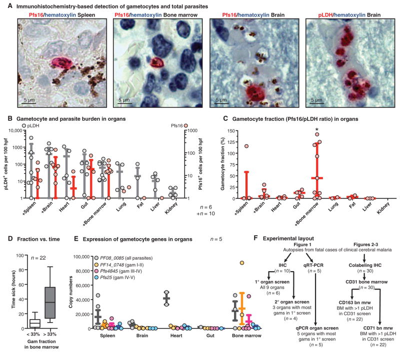
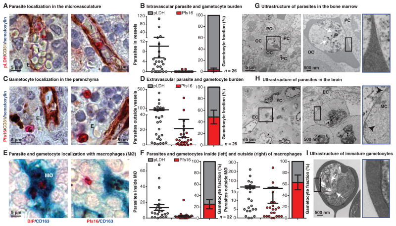
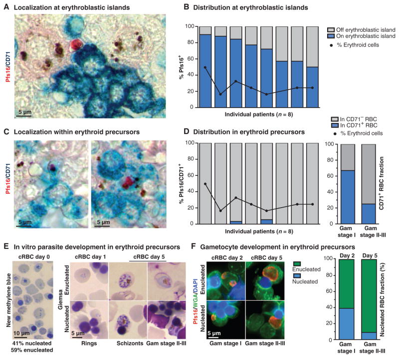
Transmission of Plasmodium falciparum malaria parasites requires formation and development of gametocytes, yet all but the most mature of these sexual parasite forms are absent from the blood circulation. We performed a systematic organ survey in pediatric cases of fatal malaria to characterize the spatial dynamics of gametocyte development in the human host. Histological studies revealed a niche in the extravascular space of the human bone marrow where gametocytes formed in erythroid precursor cells and underwent development before reentering the circulation. Accumulation of gametocytes in the hematopoietic system of human bone marrow did not rely on cytoadherence to the vasculature as does sequestration of asexual-stage parasites. This suggests a different mechanism for the sequestration of gametocytes that could potentially be exploited to block malaria transmission.


Conflict of interest statement

Competing interests: The authors declare that they have no competing interests.

Figures

Fig. 1
Fig. 1. Enrichment of developing gametocytes in the human bone marrow
(A) Detection of parasites and gametocytes across tissues. Parasite antigens (pLDH for all parasites and Pfs16 for gametocytes) were visualized with alkaline phosphatase and Fast Red substrate, and nuclei were labeled with blue hematoxylin stain. Shown are representative images of Pfs16-labeled gametocytes in the spleen, bone marrow, and brain, and of multiple pLDH-labeled sequestering parasites in the brain microvasculature. (B and C) Quantification of parasite and gametocyte load across tissues. Numbers of pLDH-positive (left axis) and Pfs16-positive (right axis) parasites were quantified per 100 high-power fields (hpf) for each organ for 6 or 10 patients (those marked with +). Dots indicate the parasite load (one dot per patient), with bars for mean and SD across patients. Gametocyte fractions (C) were calculated as a ratio of Pfs16-positive parasites to pLDH-positive parasites in 100 high-power fields from each sample, for each tissue in which at least 10 pLDH parasites were observed. Dots indicate gametocyte fraction in each patient, with bars for median and interquartile range across patients. Asterisk above graph indicates significant difference by Fisher’s exact test using categories of <33% and >33% gametocyte fraction and comparing the top five organs. (D) Distribution of time to death compared with gametocyte fraction. The time (hours) from admission to death for all patients from which the bone marrow gametocyte fraction was calculated in this study, plotted by those cases with gametocyte fractions below and above 33%. (E) Gametocyte-stage distribution across tissues by qRT-PCR. Transcript copy number for early developing (stage I/II, PF14_0748), mid developing (stage III/IV, Pfs4845), and late developing and mature gametocytes (stage IV/V, Pfs25) and the constitutive marker PF08_0085 in tissue homogenates, normalized by RNA input. RNA from five organs was analyzed from five patients. Dots indicate transcript copy number in each patient, with bars for mean and SE. (F) Summary of the autopsy cases and experimental methods used in this study. IHC, immunohistochemistry.
Fig. 2
Fig. 2. Presence of gametocytes in the hematopoietic system
(A and C) Gametocyte and parasite localization in human bone marrow. Parasite antigens were detected using alkaline phosphatase and Fast Red, endothelial cells were labeled with CD31 antibodies and detected with horseradish peroxidase (HRP) and 3,3′-diaminobenzidine (DAB) chromogen, and nuclei were labeled with blue hematoxylin stain. In representative images from one patient, ring stage (top left) and large adhering trophozoite or schizont stage (top right), parasites can be seen within bone marrow sinusoids intravascularly (I). Pfs16-labeled gametocytes can be seen in the extravascular space (E). (B and D) Quantification of bone marrow vasculature localization for Pfs16- and pLDH-positive cells. Number of pLDH- and Pfs16-labeled parasites was plotted for 50 sinusoids (intravascular) (B) or 50 high-power fields of extravascular space (D) for 30 patients (only 26 had any detectable parasites by immunohistochemistry). Mean gametocyte fractions with SE across 26 patients for intravascular (3.13 ± 3.13%) and extravascular (48.49 ± 11.74%) localization are shown. (E) Parasite and gametocyte localization within macrophages. Pfs16 and the constitutive parasite marker BIP/heat shock protein 70 were detected using alkaline phosphatase and Fast Red substrate, and macrophages (MØ) were labeled with CD163 antibodies and detected using HRP and 3,3′,5,5′-tetramethylbenzidine (TMB) substrate (turquoise). Multiple asexual schizont stages were seen within a macrophage (left), and a gametocyte is seen outside of macrophages within the bone marrow. (F) Quantification of bone marrow macrophage localization for Pfs16- and pLDH-positive cells. Shown are numbers of pLDH- and Pfs16-labeled parasites in 200 macrophages, either inside macrophages (left graph) or 50 high-power fields of extracellular space outside the macrophages (right graph) for 22 patients [those cases with more than two parasites in (D)]. Mean gametocyte fraction and SE from 22 patients for inside (24.94 ± 8.03%) and outside (63.22 ± 12.97%) macrophages. (G to I) Ultrastructural analysis of asexual and sexual parasites. Transmission electron micrograph images from the bone marrow and brain tissue samples of one patient and in vitro–derived purified stage I/II gametocytes for reference were analyzed. In the bone marrow, large knobless parasites were found in close association with multiple orthochromatic (OC) and polychromatic (PC) erythroid precursor cells (G). For comparison, sequestered asexual parasites with electron-dense knob structures (black arrowheads) and host modifications [Maurer’s clefts (MC)] were observed in brain capillaries, in close apposition to endothelial cells (EC) (H). Parasites found in the bone marrow shared key features with in vitro–derived stage I gametocytes, including multiple food vacuoles with hemozoin crystals (white arrows) and lack of knobs (I).
Fig. 3
Fig. 3. Gametocyte formation and development within the hematopoietic system
(A and C) Gametocyte localization with erythroid precursor cells in the human bone marrow. Pfs16 was detected using alkaline phosphatase and Fast Red, and erythroid precursor cells were labeled with CD71 antibodies and detected with HRP and TMB liquid substrate (turquoise). Gametocytes were seen in contact with CD71+ cells within an erythroblastic island (A) and within CD71+ cells (C). (B and D) Quantification of gametocyte localization with erythroid precursor cells. The percentage of all gametocytes quantified in 50 high-power fields that were in contact with CD71+ cells and/or the central macrophage of an erythroblastic island. The percentage of erythroid cells (as opposed to myeloid cells) is plotted for reference. Data are shown for the eight patients with the highest gametocyte load (above five gametocytes in 50 high-power fields); each bar represents one patient. A focused analysis on the gametocytes in the patient with the highest fraction of gametocytes in CD71+ cells is shown (D, right graph). The proportion of gametocytes within CD71+ cells for stage I size versus stage II to III size is shown. Gametocyte sizes as determined using ImageJ were assigned to either stage I or stage II/III on the basis of comparison with in vitro stage measurements (fig. S3). For this analysis, 15 gametocytes were measured from each category (CD71+, CD71). (E and F) In vitro parasite development in erythroid precursor cells. Cultured RBCs (cRBCs) were stained with new methylene blue to assess erythroid maturation (presence of nucleus or ribosomal RNA) on day 0 (E, left panel). Giemsa-stained smears of infected cRBCs on days 1 and 5 demonstrate asexual development and replication, as well as gametocyte development within these cells (E, right panel). For gametocyte detection, parasites were labeled with Pfs16 antibodies (red), RBC precursors were stained with wheat germ agglutinin (WGA; green), and nuclei were stained with 4′,6-diamidino-2-phenylindole (DAPI; blue). The distribution of host cell type is shown for each gametocyte-stage category, morphologically identified by size and shape of Pfs16-labeled cells.

Similar articles

Cited by

References

    1. World Health Organization. World Malaria Report 2012. World Health Organization; Geneva: 2013.
    1. Marchiafava E, Bignami A. Sulle Febbri Estivo Aumnali. E. Loescher; Torino: 1894.
    1. Thomson JG, Robertson A. The structure and development of Plasmodium falciparum gametocytes in the internal organs and peripheral circulation. Trans R Soc Trop Med Hyg. 1935;29:31–40.
    1. Farfour E, Charlotte F, Settegrana C, Miyara M, Buffet P. The extravascular compartment of the bone marrow: A niche for Plasmodium falciparum gametocyte maturation? Malar J. 2012;11:285. - PMC - PubMed
    1. Smalley ME, Abdalla S, Brown J. The distribution of Plasmodium falciparum in the peripheral blood and bone marrow of Gambian children. Trans R Soc Trop Med Hyg. 1981;75:103–105. - PubMed

Publication types

MeSH terms