. 2014 Jul 9;34(28):9235-48.
doi: 10.1523/JNEUROSCI.0561-14.2014.

Calsyntenin-1 regulates axon branching and endosomal trafficking during sensory neuron development in vivo

Affiliations

Calsyntenin-1 regulates axon branching and endosomal trafficking during sensory neuron development in vivo

Olga Y Ponomareva et al. J Neurosci. .

Abstract

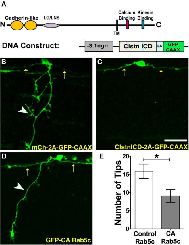
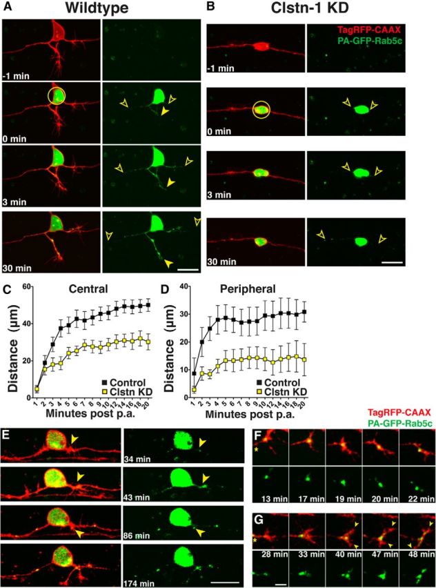
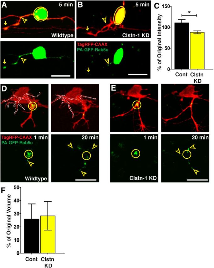
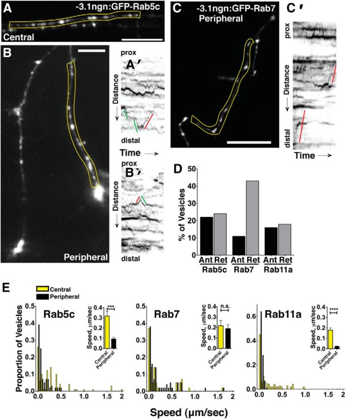
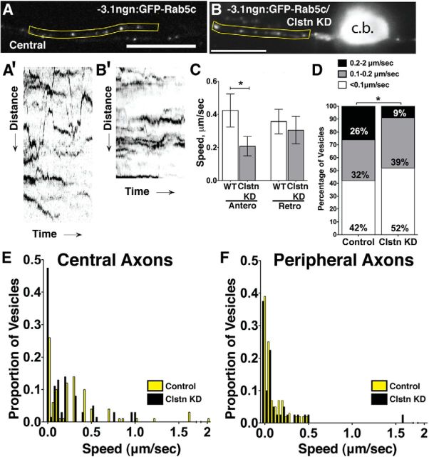
Precise regulation of axon branching is crucial for neuronal circuit formation, yet the mechanisms that control branch formation are not well understood. Moreover, the highly complex morphology of neurons makes them critically dependent on protein/membrane trafficking and transport systems, although the functions for membrane trafficking in neuronal morphogenesis are largely undefined. Here we identify a kinesin adaptor, Calsyntenin-1 (Clstn-1), as an essential regulator of axon branching and neuronal compartmentalization in vivo. We use morpholino knockdown and a Clstn-1 mutant to show that Clstn-1 is required for formation of peripheral but not central sensory axons, and for peripheral axon branching in zebrafish. We used live imaging of endosomal trafficking in vivo to show that Clstn-1 regulates transport of Rab5-containing endosomes from the cell body to specific locations of developing axons. Our results suggest a model in which Clstn-1 patterns separate axonal compartments and define their ability to branch by directing trafficking of specific endosomes.

Keywords: axon branching; calsyntenin; endosome; polarity; trafficking; zebrafish.


Figures

Figure 1.
Figure 1.
clstn1 mRNA is expressed in the developing nervous system. A–C, Lateral views of whole-mount embryos showing in situ hybridization for clstn1 mRNA at stages indicated. D, E, Dorsal (D) and lateral (E) views of expression in the head at 24 hpf. E, Inset, Dorsal expression in the eye. F, G, Lateral (F) and dorsal (G) views of the trunk at 25 hpf showing expression in the spinal cord. H, Higher-magnification view showing expression in the RB cell bodies (arrowheads) at 24 hpf. mp, Migrating primordium; te, telencephalon; tg, trigeminal ganglion; ov, otic vesicle; llg, lateral line ganglion; dien, diencephalon; mhb, midbrain-hindbrain boundary; D, dorsal; V, ventral; A, anterior; P, posterior. Scale bars, 40 μm.
Figure 2.
Figure 2.
Clstn-1 knockdown reduces branches in peripheral sensory axons. A, B, Lateral views of trigeminal ganglia labeled with anti-HNK-1 in uninjected (A) and Clstn-1 knockdown (KD) (B) embryos showing reduced branching of trigeminal axons. C, D, Lateral views of RB neurons labeled with anti-HNK-1 showing a reduction in peripheral axons in Clstn-1 KD (D) versus control MO-injected embryos (C). E, F, The focal plane of central axons in the same embryos showing no effect on the central fascicle. G–I, Single-labeled RB cells expressing GFP-CAAX show a reduction of peripheral outgrowth (H) and peripheral branching (I) in Clstn-1 KD embryos. Arrows indicate central axons; arrowheads indicate peripheral axons. Scale bars, 40 μm.
Figure 3.
Figure 3.
TALEN generation of Calsyntenin-1 mutants. A, TALENs were designed against the second exon of Clstn-1. B, clstn-1uw7and clstn-1uw8 result in nucleotide deletions with a frame shift and predicted premature stop. clstn-1uw9 has a 6 bp deletion and 9 bp insertion, which does not result in a frame shift. C, D, RB neurons labeled with HNK-1 antibody in wild-type (C) and clstn1uw7/uw7 mutant (D) embryos from incross of clstn1uw7/+ heterozygous fish. E, Quantification of axon branches crossing the horizontal myoseptum in wild-type (+/+), clstn1uw7/+ heterozygous, and clstn1uw7/uw7 homozygous embryos. **p = 0.004 (unpaired, two-tailed t test). Scale bar, 40 μm.
Figure 4.
Figure 4.
Live imaging reveals reduced peripheral branching activity in Clstn-1 knockdown. A, B, Time-lapse images (lateral views, anterior left, z-projections) from Tg(−3.1ngn1:gfp-caax) embryos injected with standard control MO (A) or Clstn-1 MO (B). Arrowheads indicate bifurcation events. C–E, Quantification of axon branching and growth rate. *p = 0.03 (unpaired, two-tailed t test). ***p = 0.0008 (unpaired, two-tailed t test). **p = 0.009 (unpaired, two-tailed t test). Scale bar, 100 μm. Time is in hours:minutes:seconds.
Figure 5.
Figure 5.
Clstn-1 intracellular domain or CA-Rab5c expression reduces peripheral RB branching. A, Schematic showing structure of Clstn-1 and a DNA construct used to express Clstn-1 ICD in RB neurons. TM, Transmembrane domain; 2A, self-cleaving peptide. B, C, Individually labeled RB neurons expressing either GFP-CAAX (B) or Clstn-1 ICD (C) showing failure of peripheral axon formation in Clstn-1 ICD-expressing cell. Arrows indicate central axons; arrowheads indicate peripheral axons. D, RB neuron expressing constitutively active Rab5c (CA-Rab5c) shows reduced branching. E, Quantification of peripheral axon tips per neuron. *p = 0.02 (unpaired, two-tailed t test). Scale bar, 40 μm.
Figure 6.
Figure 6.
Rab5 endosomes move into developing axons and Clstn-1 knockdown inhibits transport of early endosomes from the cell body into axons. A, B, Time-lapse images of PA-GFP-Rab5c-labeled endosomes in TagRFP-CAAX-labeled RB cells in control (A) and Clstn KD (B) RB neurons. Photoactivation was in the cell body (yellow circle) and spread of photoactivated Rab5c vesicles into axons was measured as the farthest vesicle from the cell body (open arrowheads). C, Quantification of Rab5 vesicle movement into central axons. Vesicles advance more slowly in Clstn-1 knockdown compared with wild-type. p = 0.0003 (two-way ANOVA). D, Rab5 vesicles move more slowly into peripheral axons in Clstn-1 knockdown embryos compared with wild-type. p = 0.03 (two-way ANOVA). E, Time-lapse images of peripheral axon initiation in neuron in which PA-GFP-Rab5 was photoactivated in the cell body. GFP-Rab5 accumulation is present at peripheral axon initiation site (arrowhead) and fills forming growth cone. F, Time-lapse images of peripheral growth cone turning. Growth cone is initially growing up, but little Rab5 enters the upper protrusion, which retracts. Growth cone redirects to area of large Rab5 accumulation. The accumulation moves into lower branch, which extends. *Axon shaft. G, Time-lapse images of bifurcating peripheral growth cone. Rab5 moves into both branches (arrowheads) of bifurcating growth cone. Scale bars: A, B, E, 20 μm; F, G, 5 μm. Time is minutes after photoactivation.
Figure 7.
Figure 7.
Clstn-1 knockdown reduces Rab5 vesicle delivery to branch points. A, Endosomes accumulate at branch points (open yellow arrowheads) and in growth cones (arrows) of peripheral axons in control embryos. B, Clstn-1 knockdown embryos show reduced accumulations at branch points. C, Quantification of GFP accumulation intensity change at branch points (intensity at 30 min after activation divided by intensity at 3 min after activation). *p = 0.03 (unpaired, two-tailed t test). D, E, Photoactivation in branch point (yellow circle) of control neuron (D) or Clstn-1 KD neuron (E). Signal is largely dispersed from the branch points by 20 min after PA. Arrowheads indicate vesicles that moved from branch point into axon branches. Left branch in D is outlined for clarity. F, Quantification of change in vesicle accumulation at branch points (volume at 20 min divided by volume at 2 min) shows no difference between control and Clstn-1 KD (p = 0.885). Scale bars, 20 μm. Time is minutes after photoactivation.
Figure 8.
Figure 8.
Characterization of endosome dynamics in RB axons. A, B, GFP-Rab5c-labeled endosomes in central (A) and peripheral (B) axons. A′, Kymograph of yellow region in A with lines indicating anterograde (green) or retrograde (red) movement, with reference to the cell body. prox, Proximal. B′, Kymograph of peripheral Rab5c endosomes in yellow region from B. C, Peripheral RB with GFP-Rab7-labeled late endosomes. C′, Kymograph of yellow region in C showing retrograde (red) movements. D, Directionality of GFP-Rab5c-, GFP-Rab7-, and GFP-Rab11-labeled endosomes in RB cells. Vesicles with no net movement during the imaging period are not shown in the graph. E, Speed distributions of GFP-Rab5c-, GFP-Rab7-, and GFP-Rab11-labeled endosomes in central (yellow) and peripheral (black) axons. Insets, Mean speeds in central and peripheral axons. ***p = 0.001 (unpaired, two-tailed t test). ****p < 0.0001 (unpaired, two-tailed t test). n.s., Not significant. Scale bar, 20 μm.
Figure 9.
Figure 9.
Clstn-1 knockdown reduces early endosomal speeds. A, B, Single frames from time-lapse movies of GFP-labeled Rab5c in central RB axons of control (A) and Clstn-1 KD (B) embryos at 24 hpf. c.b., Cell body. A′, B′, Kymographs from yellow regions of A and B of vesicle movement in control (A′) and Clstn-1 knockdown (B′) central RB axons, showing reduced movement in Clstn-1 knockdown embryos. C, Speeds of vesicles moving in the anterograde directions were significantly reduced. *p = 0.05 (unpaired, two-tailed t test). D, Percentage of vesicles moving at specific speeds shows fewer early endosomes traveling at fast speeds in central axons of Clstn-1 knockdown embryos. *p = 0.03 (χ2 test). E, F, Speed distributions of endosomes in central (E) and peripheral (F) axons of control (yellow) and Clstn-1 knockdown (black) embryos.

Similar articles

Cited by

References

    1. Andersen E, Asuri N, Clay M, Halloran M. Live imaging of cell motility and actin cytoskeleton of individual neurons and neural crest cells in zebrafish embryos. J Vis Exp. 2010;36:1726. doi: 10.3791/1726. - DOI - PMC - PubMed
    1. Andersen EF, Asuri NS, Halloran MC. In vivo imaging of cell behaviors and F-actin reveals LIM-HD transcription factor regulation of peripheral versus central sensory axon development. Neural Dev. 2011;6:27. doi: 10.1186/1749-8104-6-27. - DOI - PMC - PubMed
    1. Araki Y, Tomita S, Yamaguchi H, Miyagi N, Sumioka A, Kirino Y, Suzuki T. Novel cadherin-related membrane proteins, Alcadeins, enhance the X11-like protein-mediated stabilization of amyloid beta-protein precursor metabolism. J Biol Chem. 2003;278:49448–49458. doi: 10.1074/jbc.M306024200. - DOI - PubMed
    1. Araki Y, Kawano T, Taru H, Saito Y, Wada S, Miyamoto K, Kobayashi H, Ishikawa HO, Ohsugi Y, Yamamoto T, Matsuno K, Kinjo M, Suzuki T. The novel cargo Alcadein induces vesicle association of kinesin-1 motor components and activates axonal transport. EMBO J. 2007;26:1475–1486. doi: 10.1038/sj.emboj.7601609. - DOI - PMC - PubMed
    1. Ascaño M, Richmond A, Borden P, Kuruvilla R. Axonal targeting of Trk receptors via transcytosis regulates sensitivity to neurotrophin responses. J Neurosci. 2009;29:11674–11685. doi: 10.1523/JNEUROSCI.1542-09.2009. - DOI - PMC - PubMed

Publication types

Substances

LinkOut - more resources