. 2014 Jul 9;34(28):9249-60.
doi: 10.1523/JNEUROSCI.3979-13.2014.

The role of the hypothalamic paraventricular nucleus and the organum vasculosum lateral terminalis in the control of sodium appetite in male rats

Affiliations

The role of the hypothalamic paraventricular nucleus and the organum vasculosum lateral terminalis in the control of sodium appetite in male rats

Laura A Grafe et al. J Neurosci. .

Abstract

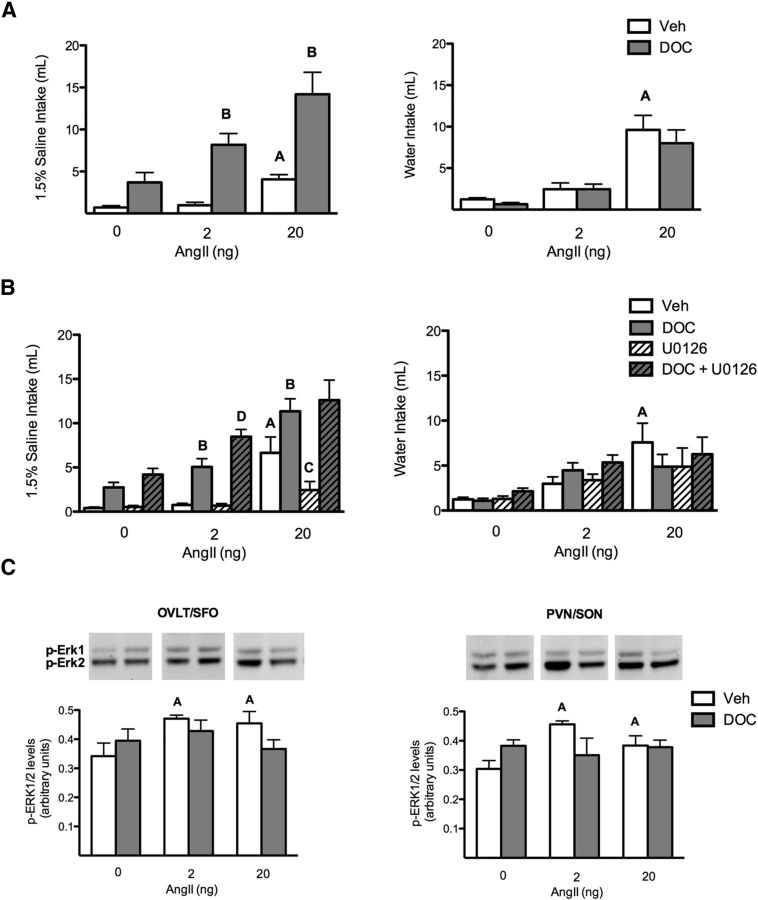
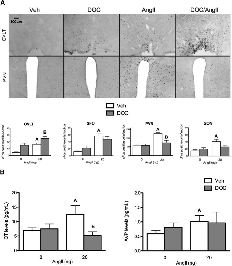
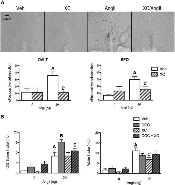
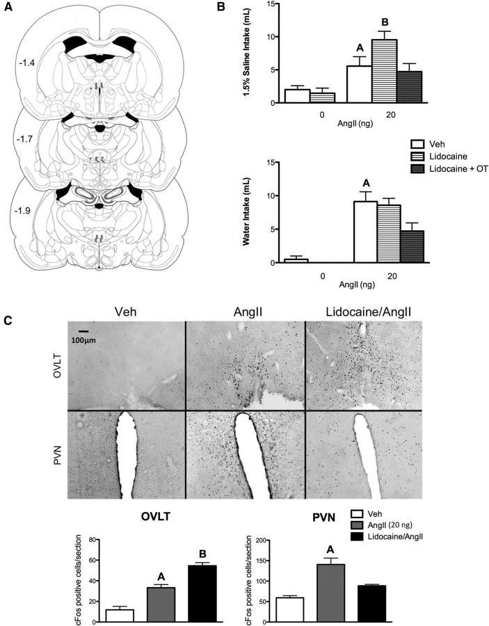
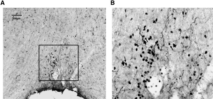
Angiotensin II (AngII) and aldosterone cooperate centrally to produce a robust sodium appetite. The intracellular signaling and circuitry that underlie this interaction remain unspecified. Male rats pretreated with both deoxycorticosterone (DOC; a synthetic precursor of aldosterone) and central AngII exhibited a marked sodium intake, as classically described. Disruption of inositol trisphosphate signaling, but not extracellular-regulated receptor kinase 1 and 2 signaling, prevented the cooperativity of DOC and AngII on sodium intake. The pattern of expression of the immediate early gene product cFos was used to identify key brain regions that may underlie this behavior. In the paraventricular nuclei (PVN) of the hypothalamus, DOC pretreatment diminished both AngII-induced cFos induction and neurosecretion of oxytocin, a peptide expressed in the PVN. Conversely, in the organum vasculosum lateral terminalis (OVLT), DOC pretreatment augmented cFos expression. Immunohistochemistry identified a substantial presence of oxytocin fibers in the OVLT. In addition, when action potentials in the PVN were inhibited with intraparenchymal lidocaine, AngII-induced sodium ingestion was exaggerated. Intriguingly, this treatment also increased the number of neurons in the OVLT expressing AngII-induced cFos. Collectively, these results suggest that the behavioral cooperativity between DOC and AngII involves the alleviation of an inhibitory oxytocin signal, possibly relayed directly from the PVN to the OVLT.

Keywords: aldosterone; angiotensin; oxytocin; receptor signaling; sodium appetite.


Figures

Figure 1.
Figure 1.
Erk1/2 is not necessary for the DOC/AngII potentiation of sodium appetite. A, Bar graphs illustrating the potentiation of AngII-induced sodium but not water intake by DOC (n = 12/group). B, Bar graphs illustrating the effect of blocking ERK1/2 activation on DOC and AngII-induced sodium and water intake (n = 18/group). DOC potentiated 2 and 20 ng AngII-induced sodium intake. This behavior was not reduced by the MEK inhibitor U0126; however, U0126 reduced sodium intake with 20 ng AngII alone. AngII-induced water intake was not potentiated by DOC. C, Bar graphs illustrating phosphorylated ERK1/2 levels in the OVLT/SFO and PVN/SON after either oil or DOC pretreatment followed by intracerebroventricular treatments with 2 or 20 ng AngII (n = 6/group). AngII treatment induced significant ERK1/2 phosphorylation compared with vehicle in both sets of brain regions. DOC pretreatment did not enhance this activation. Representative Western blot images of ERK1/2 phosphorylation are shown above each quantified bar. For the sake of clarity, only specific statistical differences are highlighted. “A” indicates AngII is different from vehicle; “B” indicates DOC/AngII is different from AngII; “C” indicates U0126/AngII is different from AngII; and “D” indicates U0126/DOC/AngII is different from DOC/AngII, p < 0.05. Veh, Vehicle.
Figure 2.
Figure 2.
DOC pretreatment enhanced AngII-induced OVLT activation and reduced PVN activity and oxytocin (OT) release. A, Bar graphs illustrating cFos cell counts in different brain areas (OVLT, SFO, PVN, and SON) after either vehicle or DOC pretreatment followed by intracerebroventricular vehicle or 20 ng AngII (n = 6/group). In the OVLT, each treatment condition induces cFos immunostaining compared with vehicle; DOC/AngII induced additional cFos immunostaining. In the SFO, both AngII alone and DOC/AngII induced more cFos staining than either vehicle or DOC alone. cFos labeling in the PVN was increased by AngII, but DOC reduced the AngII-induced cFos expression to control levels. In the SON, AngII induced cFos staining. Representative images of the OVLT and PVN (coronal plane, 10×) in each treatment condition are shown above the bar graphs. Representative images of SFO and SON are not shown, but similar data were seen. B, Bar graphs illustrating oxytocin and AVP plasma levels after either vehicle or DOC pretreatment followed by intracerebroventricular vehicle or 20 ng AngII (n = 8/group). AngII treatment increased oxytocin levels, and this effect was reduced by DOC pretreatment. However, AngII treatment elevated AVP levels, regardless of pretreatment. For the sake of clarity, only specific statistical differences are highlighted. “A” indicates AngII is different from vehicle; “B” indicates DOC/AngII is different from AngII. Veh, Vehicle.
Figure 3.
Figure 3.
The IP3 receptor is required for DOC/AngII potentiation of sodium appetite. A, Bar graphs illustrating the level of cFos expression in the OVLT and SFO after vehicle, XC, 20 ng AngII, or XC plus 20 ng AngII intracerebroventricularly (n = 6/group). Representative images of cFos staining of OVLT coronal sections (10×) in each treatment group are shown above the graphs. Representative images of the SFO are not shown, but similar data were seen. AngII induced cFos staining in both the OVLT and SFO, and the effect was reduced by XC. B, Bar graphs illustrating the effect of XC on DOC and 20 ng AngII induced sodium and water intake (n = 10/group). DOC potentiates AngII-induced sodium intake; this effect was impaired by the IP3 receptor inhibitor XC. XC did not affect sodium intake induced by AngII alone. AngII-induced water intake was reduced by XC. For the sake of clarity, only specific statistical differences are highlighted. “A” indicates AngII is different from vehicle; “B” indicates DOC/AngII is different from AngII; “C” indicates XC/AngII is different from AngII; “D” indicates XC/DOC/AngII is different from DOC/AngII; p < 0.05. Veh, vehicle.
Figure 4.
Figure 4.
PVN inactivation enhances AngII-induced sodium appetite. A, Illustration of rat coronal brain slices depicting bilateral guide cannula placements (filled circles) above the PVN for lidocaine administration. Each pair of dots represents data from one animal. B, Bar graphs illustrating the effect of intra-PVN lidocaine on 20 ng AngII-induced sodium and water intake (n = 10/group). Lidocaine-treated animals increased their AngII-induced sodium intake, but not water intake, mimicking the effects of DOC. C, Bar graphs illustrating cFos expression in the OVLT and PVN after vehicle, lidocaine, 20 ng AngII, or lidocaine plus 20 ng AngII intracerebroventricularly (n = 6/group). Representative images of cFos staining of OVLT and PVN coronal sections (10×) in each treatment group are shown above the graphs. AngII-induced cFos immunostaining was enhanced by lidocaine in the OVLT and reduced in the PVN. For the sake of clarity, only specific statistical differences are highlighted. “A” indicates AngII is different from vehicle; “B” indicates lidocaine/AngII is different from AngII. Veh, Vehicle.
Figure 5.
Figure 5.
Oxytocin fibers are closely associated with AngII-induced cFos in the dorsal cap of the OVLT. A, Representative image of oxytocin and cFos staining of an OVLT coronal section (10×) in an AngII-treated animal. B, Magnified image of the dorsal cap of the OVLT (from the box drawn in A). Thin and thick arrows indicate certain cFos nuclei and OVLT fibers, respectively, that are juxtaposed throughout the dorsal cap.

Similar articles

Cited by

References

    1. Akaishi T, Negoro H, Kobayasi S. Responses of paraventricular and supraoptic units to angiotensin II, sar1-ile8-angiotensin II and hypertonic NaCl administered into the cerebral ventricle. Brain Res. 1980;188:499–511. doi: 10.1016/0006-8993(80)90048-7. - DOI - PubMed
    1. Andersson B, Leksell LG, Lishajko F. Perturbations in fluid balance induced by medially placed forebrain lesions. Brain Res. 1975;99:261–275. doi: 10.1016/0006-8993(75)90028-1. - DOI - PubMed
    1. Beresford MJ, Fitzsimons JT. Intracerebroventricular angiotensin II-induced thirst and sodium appetite in rat are blocked by the AT1 receptor antagonist, losartan (Dup753), but not by the AT2 antagonist, CGP 42112A. Exp Physiol. 1992;77:761–764. - PubMed
    1. Bisley JW, Rees SM, McKinley MJ, Hards DK, Oldfield BJ. Identification of osmoresponsive neurons in the forebrain of the rat: A fos study at the ultrastructural level. Brain Res. 1996;720:25–34. doi: 10.1016/0006-8993(96)00079-0. - DOI - PubMed
    1. Bisset GW, Clark BJ, Errington ML. The hypothalamic neurosecretory pathways for the release of oxytocin and vasopressin in the cat. J Physiol. 1971;217:111–131. - PMC - PubMed

Publication types

LinkOut - more resources