. 2014 Jul 9;34(28):9364-76.
doi: 10.1523/JNEUROSCI.4787-13.2014.

Synucleins regulate the kinetics of synaptic vesicle endocytosis

Affiliations

Synucleins regulate the kinetics of synaptic vesicle endocytosis

Karina J Vargas et al. J Neurosci. .

Abstract

Genetic and pathological studies link α-synuclein to the etiology of Parkinson's disease (PD), but the normal function of this presynaptic protein remains unknown. α-Synuclein, an acidic lipid binding protein, shares high sequence identity with β- and γ-synuclein. Previous studies have implicated synucleins in synaptic vesicle (SV) trafficking, although the precise site of synuclein action continues to be unclear. Here we show, using optical imaging, electron microscopy, and slice electrophysiology, that synucleins are required for the fast kinetics of SV endocytosis. Slowed endocytosis observed in synuclein null cultures can be rescued by individually expressing mouse α-, β-, or γ-synuclein, indicating they are functionally redundant. Through comparisons to dynamin knock-out synapses and biochemical experiments, we suggest that synucleins act at early steps of SV endocytosis. Our results categorize α-synuclein with other familial PD genes known to regulate SV endocytosis, implicating this pathway in PD.

Keywords: AP180; Parkinson's disease; endocytosis; membrane bending; presynaptic; synaptobrevin.


Figures

Figure 1.
Figure 1.
Synucleins regulate SV endocytosis. a, Average representative traces of spH fluorescence in wild-type and αβγ-Syn−/− neurons. Neurons were stimulated three times (100 AP, 300 AP, and 600 AP) at 20 Hz with an interval of 2 min between each stimulation. Average time constants of decay (τ) for wild-type = 15.6 s, 13.3 s, 17.8 s, respectively, for αβγ-Syn−/− = 36.1 s, 35.6 s, 27.6 s, respectively; n = 6 neurons/3 independent experiments for wild-type and n = 4 neurons/3 independent experiments for αβγ-Syn−/−. Inset, The 100 AP, 20 Hz stimulation trace scaled to peak fluorescence, showing that the decay kinetics is slowed in αβγ-Syn−/− neurons mice b, c, spH trace of wild-type and αβγ-Syn−/− neurons stimulated in the presence (purple) or absence (blue) of bafilomycin followed by NH4Cl (n = 9 wild-type neurons/4 experiments and 12 αβγ-Syn−/− neurons/6 experiments). The variance in the wild-type trace is greater than αβγ-Syn−/− post stimulus, therefore, this portion of the trace was not analyzed further. d, e, spH responses in presence and absence of bafilomycin (bafilo) during the first 5 s of stimulation were subtracted to obtain endocytic rate constant (orange). f, g, The average exocytic (f) and endocytic (g) rate calculated from d and e. h, Average maximum fluorescence after exposure to 50 mm NH4Cl; *p < 0.05.
Figure 2.
Figure 2.
Mouse synucleins are functionally redundant a, Average spH fluorescence normalized to the maximum fluorescence value in wild-type (blue squares) and αβγ-Syn−/− (green triangles) neurons. (n = 16 wild-type and 15 αβγ-Syn−/− neurons/3 independent experiments). b, Average time constants of decay (τ) of spH signal after stimulation calculated for wild-type, αβγ-Syn−/−, and αβγ-Syn−/− neurons transfected with mouse α-Syn (yellow), β-Syn (brown), and γ-Syn (red). t test ***p < 0.001 wild-type versus αβγ-Syn−/−; wild-type versus synuclein rescues; αβγ-Syn−/− versus synuclein rescues significant *p < 0.05, **p < 0.01.
Figure 3.
Figure 3.
CT-HRP labeling reveals an endocytic deficit in αβγ-Syn−/− synapses. a, b, Representative electron micrographs of wild-type and αβγ-Syn−/− hippocampal neurons undergoing the following stimulation protocol: Tyrode's buffer for 5′ (Rest), 90 mm KCl for 2′ (K+), followed by Tyrode's buffer for 10′ (Recovery) in the constant presence of CT-HRP. Red arrowhead, CT-HRP-labeled SV and red arrow, endosomes. c, d, Fraction of CT-HRP labeled (c) endosomes and (d) SVs in wild-type and αβγ-Syn−/− neurons during steps of the stimulation protocol; n = 125–238 synapses (wild-type: Rest = 238, K+ = 166, Recovery 173; αβγ-Syn−/−: Rest = 176, K+ = 125, Recovery = 180)/6 experiments. *p < 0.05, ***p < 0.001. Scale bars: b, 400 nm; b, inset, 200 nm.
Figure 4.
Figure 4.
Impaired synaptic depletion and recovery in synuclein null brains. a, Representative synaptic responses generated by repetitive trains (300 stimuli, 15 Hz) in wild-type and αβγ-Syn−/− mice. Traces are averages of individual animals (n = 24/5 wild-type and 20/5 αβγ-Syn−/− slices/animal). b, Summary data showing amplitudes of fEPSPs in depletion train. Responses from each slice were normalized to the peak fEPSP amplitude in the train. Inset calibration: 20% of fEPSP amplitude, 500 ms. c, Individual fEPSPs immediately after the depletion train showed impaired recovery in αβγ-Syn−/− slices. The recovery paradigm was initiated 250 ms following the depletion train. Traces are averages of individual animals (4 slices/animal). d, Summary of synaptic recovery in wild-type and αβγ-Syn−/− mice. Responses from each slice were normalized to the first fEPSP in the depletion train. Inset, Slope values represent percentage baseline recovered per stimulus number. Note slower recovery rate in αβγ-Syn−/− slices.
Figure 5.
Figure 5.
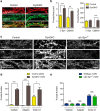
Synucleins act prior to the action of dynamins. a, b, Colocalization of α-synuclein and clathrin in DynKO neurons. a, Representative images and (b) quantification of α-synuclein and clathrin fluorescence puncta size and number (> 1 μm2) in control and DynKO neurons. Scale bar: a, 7 μm. c, Representative immunofluorescence images of clathrin, α-adaptin, and endophilin A1 in Control, DynDKO, and αβγ-Syn−/− neurons. Scale bar, 7 μm. d, e, Quantification of clathrin, α-adaptin, and endophilin A1 fluorescence puncta (> 1 μm2) in control and DynDKO neurons (d), and in wild-type and αβγ-Syn−/− neurons (e) DynDKO; n = 12–20 images/4 experiments, except for endophilin A1 n = 6–16 images/2 experiments. αβγ-Syn−/− 12 images/6 experiments.
Figure 6.
Figure 6.
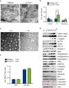
Lack of enrichment of CCVs in αβγ-Syn−/− synapses. a, Representative electron microscopic images used for quantification of CCVs in wild-type and αβγ-Syn−/− synapses. Electron microscopic images showing CCVs in wild-type and αβγ-Syn−/− synapses after high KCl stimulation. Scale bars: 400 nm; inset, 200 nm. b, CCV number in wild-type (blue) and αβγ-Syn−/− (green) at rest, after 90 s of 90 mm KCl stimulation, and 10 min of subsequent recovery. CCV numbers in αβγ-Syn−/− are not significantly different from wild-type, though there is a tendency for increased CCV upon KCl stimulation; n = 6 coverslips/3 experiments; 50–100 synapses each condition. c, Electron microscopic images of CCV and SVs derived from a biochemical fractionation of wild-type brains (Maycox et al., 1992), confirming the high purity of these fractions. d, Subcellular fractionation of wild-type brains showing the initial homogenate (H), postnuclear pellet (P1), cytosol (S2), crude synaptosomes (P2), SVs, and CCVs. Immunodetection of CCV components, SV proteins, and endocytic and exocytic proteins as well as α- and β-synuclein. The enrichment of integral CCV proteins and replication of known fractionation patterns for other synaptic proteins corroborates the success of the fractionation. Similar results were obtained in three independent experiments. e, Total acidic brain lipids were extracted and quantified as described by Volpicelli-Daley et al. (2010). PI(4)P and PI(4, 5)P2 amounts were normalized to the total amount of lipids. The levels of all phosphoinositides are similar in wild-type (blue bars) and synuclein null (αβγ-Syn−/−; green bars) brains; n = 6 brains/genotype.
Figure 7.
Figure 7.
Synucleins act at early steps in SV endocytosis. a, Quantification of presynaptic proteins in synaptosomes isolated from wild-type and αβγ-Syn−/− brains. b, Subcellular fractionation of wild-type and αβγ-Syn−/− brains showing initial homogenate (H), crude synaptosomes (P2′), fractions LS1 and LP1 en route to purify synaptic cytosol LS2, SVs, and synaptic plasma membrane (SPM), myelin, and mitochondria. Immunoblotting of amphiphysin as a synaptic cytosolic protein and synaptogyrin 1 as a SV protein to confirm purity of fractions. Purified SVs were subject to iTRAQ analysis (n = 3 independent experiments), and synaptobrevin 2 levels were found to be decreased in αβγ-Syn−/− SVs and confirmed by quantitative immunoblotting. c, Immunoprecipitations using AP180 and pre-immune serum from wild-type (WT) and αβγ-Syn−/− (KO) synaptosomes without (−) or by KCl stimulation (+). d, Quantification of immunoblots such as shown in c. From three independent experiments.
Figure 8.
Figure 8.
Model of synucleins function during SV endocytosis. Based on our results, we propose that synucleins act during early steps on SV endocytosis, which involve membrane bending and cargo selection. Horizontal lines indicate duration of action of the given endocytic protein. The blue color denotes PI(4,5)P2 levels.

References

    1. Anwar S, Peters O, Millership S, Ninkina N, Doig N, Connor-Robson N, Threlfell S, Kooner G, Deacon RM, Bannerman DM, Bolam JP, Chandra SS, Cragg SJ, Wade-Martins R, Buchman VL. Functional alterations to the nigrostriatal system in mice lacking all three members of the synuclein family. J Neurosci. 2011;31:7264–7274. doi: 10.1523/JNEUROSCI.6194-10.2011. - DOI - PMC - PubMed
    1. Balaji J, Ryan TA. Single-vesicle imaging reveals that synaptic vesicle exocytosis and endocytosis are coupled by a single stochastic mode. Proc Natl Acad Sci U S A. 2007;104:20576–20581. doi: 10.1073/pnas.0707574105. - DOI - PMC - PubMed
    1. Ben Gedalya T, Loeb V, Israeli E, Altschuler Y, Selkoe DJ, Sharon R. Alpha-synuclein and polyunsaturated fatty acids promote clathrin-mediated endocytosis and synaptic vesicle recycling. Traffic. 2009;10:218–234. doi: 10.1111/j.1600-0854.2008.00853.x. - DOI - PMC - PubMed
    1. Bendor JT, Logan TP, Edwards RH. The function of alpha-synuclein. Neuron. 2013;79:1044–1066. doi: 10.1016/j.neuron.2013.09.004. - DOI - PMC - PubMed
    1. Blondeau F, Ritter B, Allaire PD, Wasiak S, Girard M, Hussain NK, Angers A, Legendre-Guillemin V, Roy L, Boismenu D, Kearney RE, Bell AW, Bergeron JJ, McPherson PS. Tandem MS analysis of brain clathrin-coated vesicles reveals their critical involvement in synaptic vesicle recycling. Proc Natl Acad Sci U S A. 2004;101:3833–3838. doi: 10.1073/pnas.0308186101. - DOI - PMC - PubMed

Publication types