

DIPA-family coiled-coils bind conserved isoform-specific head domain of p120-catenin family: potential roles in hydrocephalus and heterotopia
- PMID: 25009281
- PMCID: PMC4148249
- DOI: 10.1091/mbc.E13-08-0492
DIPA-family coiled-coils bind conserved isoform-specific head domain of p120-catenin family: potential roles in hydrocephalus and heterotopia
Abstract
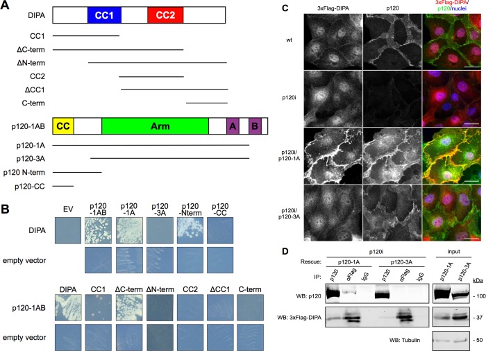
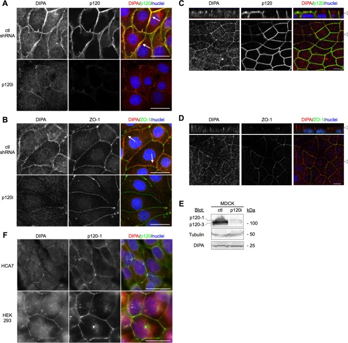
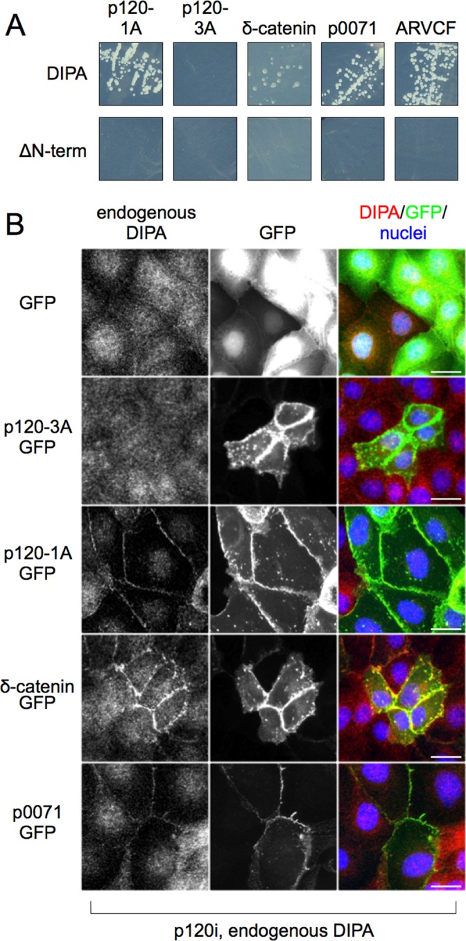
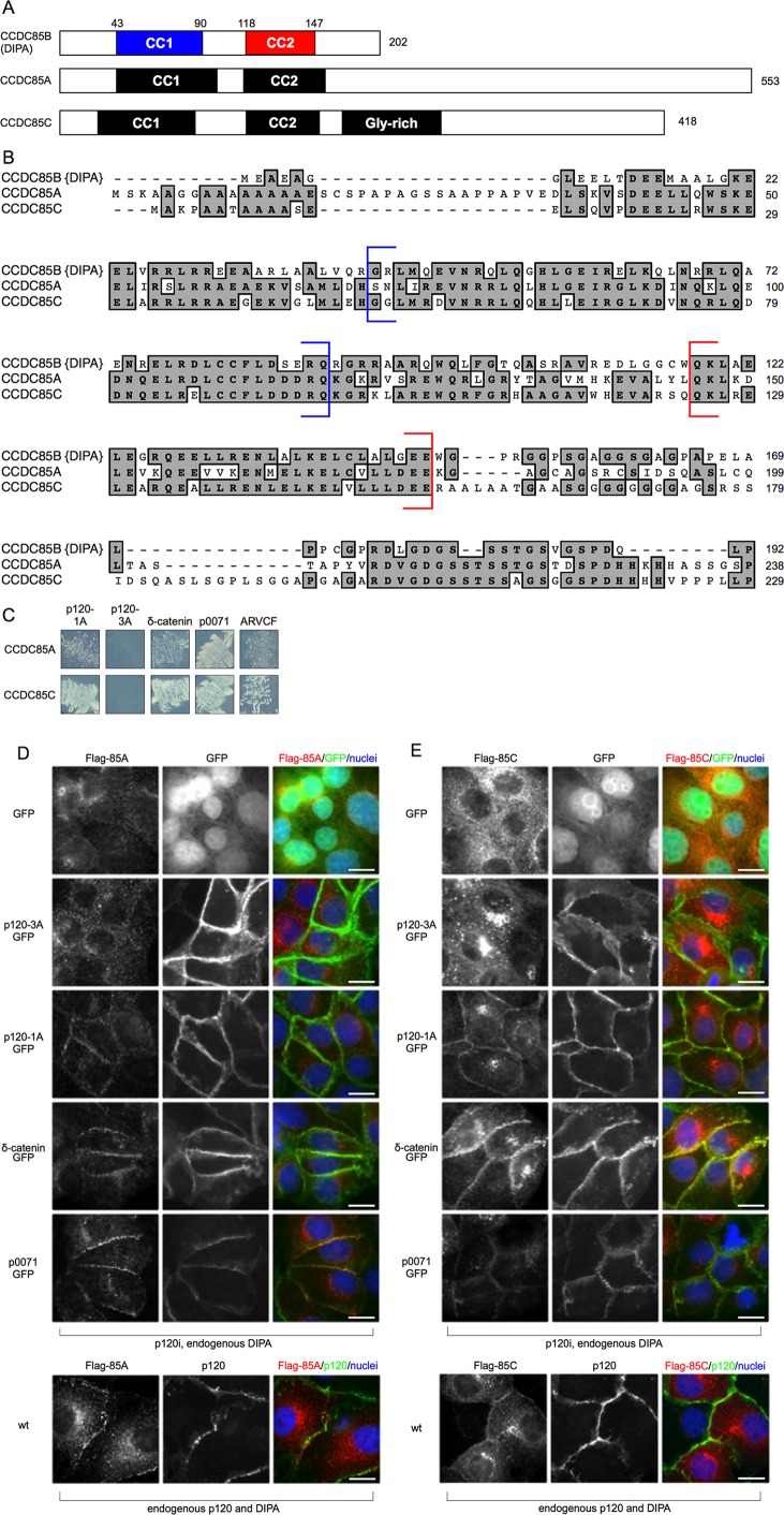
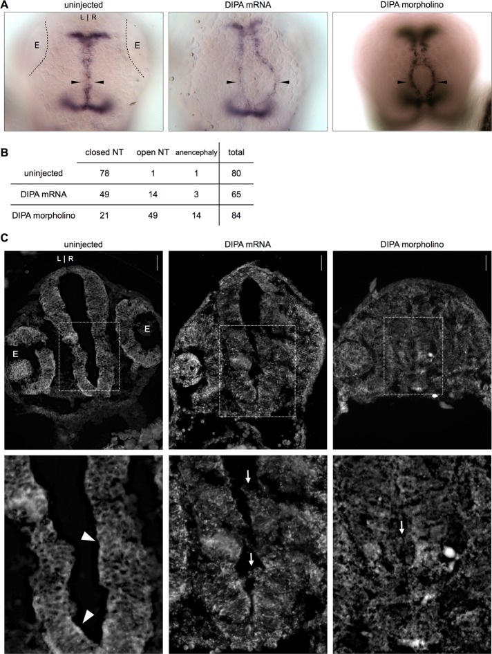
p120-catenin (p120) modulates adherens junction (AJ) dynamics by controlling the stability of classical cadherins. Among all p120 isoforms, p120-3A and p120-1A are the most prevalent. Both stabilize cadherins, but p120-3A is preferred in epithelia, whereas p120-1A takes precedence in neurons, fibroblasts, and macrophages. During epithelial-to-mesenchymal transition, E- to N-cadherin switching coincides with p120-3A to -1A alternative splicing. These isoforms differ by a 101-amino acid "head domain" comprising the p120-1A N-terminus. Although its exact role is unknown, the head domain likely mediates developmental and cancer-associated events linked to p120-1A expression (e.g., motility, invasion, metastasis). Here we identified delta-interacting protein A (DIPA) as the first head domain-specific binding partner and candidate mediator of isoform 1A activity. DIPA colocalizes with AJs in a p120-1A- but not 3A-dependent manner. Moreover, all DIPA family members (Ccdc85a, Ccdc85b/DIPA, and Ccdc85c) interact reciprocally with p120 family members (p120, δ-catenin, p0071, and ARVCF), suggesting significant functional overlap. During zebrafish neural tube development, both knockdown and overexpression of DIPA phenocopy N-cadherin mutations, an effect bearing functional ties to a reported mouse hydrocephalus phenotype associated with Ccdc85c. These studies identify a novel, highly conserved interaction between two protein families that may participate either individually or collectively in N-cadherin-mediated development.
© 2014 Markham et al. This article is distributed by The American Society for Cell Biology under license from the author(s). Two months after publication it is available to the public under an Attribution–Noncommercial–Share Alike 3.0 Unported Creative Commons License (http://creativecommons.org/licenses/by-nc-sa/3.0).
Figures
Similar articles
-
Monoclonal antibodies to DIPA: a novel binding partner of p120-catenin isoform 1.Hybridoma (Larchmt). 2012 Aug;31(4):246-54. doi: 10.1089/hyb.2012.0009. Hybridoma (Larchmt). 2012. PMID: 22894777 Free PMC article.
-
N-terminal 1-54 amino acid sequence and Armadillo repeat domain are indispensable for P120-catenin isoform 1A in regulating E-cadherin.PLoS One. 2012;7(5):e37008. doi: 10.1371/journal.pone.0037008. Epub 2012 May 16. PLoS One. 2012. PMID: 22615871 Free PMC article.
-
The molecular evolution of the p120-catenin subfamily and its functional associations.PLoS One. 2010 Dec 31;5(12):e15747. doi: 10.1371/journal.pone.0015747. PLoS One. 2010. PMID: 21209830 Free PMC article.
-
Functions of p120ctn in development and disease.Front Biosci (Landmark Ed). 2012 Jan 1;17(2):760-83. doi: 10.2741/3956. Front Biosci (Landmark Ed). 2012. PMID: 22201773 Review.
-
p120 catenin: an essential regulator of cadherin stability, adhesion-induced signaling, and cancer progression.Prog Mol Biol Transl Sci. 2013;116:409-32. doi: 10.1016/B978-0-12-394311-8.00018-2. Prog Mol Biol Transl Sci. 2013. PMID: 23481205 Free PMC article. Review.
Cited by
-
Single-Cell Transcriptomics Reveals Regulators of Neuronal Migration and Maturation During Brain Development.J Exp Neurosci. 2018 Mar 8;12:1179069518760783. doi: 10.1177/1179069518760783. eCollection 2018. J Exp Neurosci. 2018. PMID: 29551912 Free PMC article.
-
Beyond β-catenin: prospects for a larger catenin network in the nucleus.Nat Rev Mol Cell Biol. 2016 Jan;17(1):55-64. doi: 10.1038/nrm.2015.3. Epub 2015 Nov 18. Nat Rev Mol Cell Biol. 2016. PMID: 26580716 Free PMC article. Review.
-
Getting to the core of cadherin complex function in Caenorhabditis elegans.F1000Res. 2015 Dec 18;4:F1000 Faculty Rev-1473. doi: 10.12688/f1000research.6866.1. eCollection 2015. F1000Res. 2015. PMID: 26918136 Free PMC article. Review.
-
Genetic and molecular mechanisms of hydrocephalus.Front Mol Neurosci. 2025 Jan 7;17:1512455. doi: 10.3389/fnmol.2024.1512455. eCollection 2024. Front Mol Neurosci. 2025. PMID: 39839745 Free PMC article. Review.
-
An instructive role for C. elegans E-cadherin in translating cell contact cues into cortical polarity.Nat Cell Biol. 2015 Jun;17(6):726-35. doi: 10.1038/ncb3168. Epub 2015 May 4. Nat Cell Biol. 2015. PMID: 25938815 Free PMC article.
References
-
- Aho S, Rothenberger K, Uitto J. Human p120ctn catenin: tissue-specific expression of isoforms and molecular interactions with BP180/type XVII collagen. J Cell Biochem. 1999;73:390–399. - PubMed
-
- Anastasiadis PZ, Moon SY, Thoreson MA, Mariner DJ, Crawford HC, Zheng Y, Reynolds AB. Inhibition of RhoA by p120 catenin. Nat Cell Biol. 2000;2:637–644. - PubMed
-
- Anastasiadis PZ, Reynolds AB. The p120 catenin family: complex roles in adhesion, signaling and cancer. J Cell Sci. 2000;113:1319–1334. - PubMed
-
- Bezy O, Elabd C, Cochet O, Petersen RK, Kristiansen K, Dani C, Ailhaud G, Amri E-Z. Delta-interacting protein A, a new inhibitory partner of CCAAT/enhancer-binding protein beta, implicated in adipocyte differentiation. J Biol Chem. 2005;280:11432–11438. - PubMed
Publication types
MeSH terms
Substances
Grants and funding
- UL1 TR000445/TR/NCATS NIH HHS/United States
- R01 GM107079/GM/NIGMS NIH HHS/United States
- R01 GM102524/GM/NIGMS NIH HHS/United States
- P50-CA95103/CA/NCI NIH HHS/United States
- T32 GM007347/GM/NIGMS NIH HHS/United States
- T32 CA009592/CA/NCI NIH HHS/United States
- R01CA55724/CA/NCI NIH HHS/United States
- R01 HD054534/HD/NICHD NIH HHS/United States
- R01HD054534/HD/NICHD NIH HHS/United States
- R01 GM052112/GM/NIGMS NIH HHS/United States
- P50 CA095103/CA/NCI NIH HHS/United States
- K01 DK092320/DK/NIDDK NIH HHS/United States
- R01 CA046413/CA/NCI NIH HHS/United States
- T32 GM07347/GM/NIGMS NIH HHS/United States
- R01CA111947/CA/NCI NIH HHS/United States
- P30-CA11947/CA/NCI NIH HHS/United States
LinkOut - more resources
Full Text Sources
Other Literature Sources
Medical
Molecular Biology Databases
Research Materials
