

Right hepatectomy for giant cavernous hemangioma with diffuse hemangiomatosis around Glisson's capsule
- PMID: 25009410
- PMCID: PMC4081710
- DOI: 10.3748/wjg.v20.i25.8312
Right hepatectomy for giant cavernous hemangioma with diffuse hemangiomatosis around Glisson's capsule
Abstract
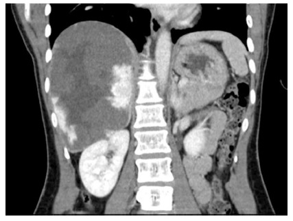
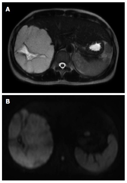
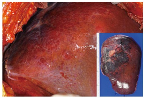
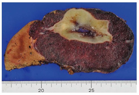
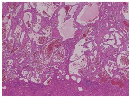
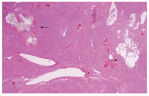
Diffuse liver hemangiomatosis with giant cavernous hemangioma in adult is extremely rare. A 35 year-old woman presented to hospital with main complaint of epigastric pain and abdominal fullness. An enhanced computed tomography scan revealed a massive liver tumor in right lobe about 150 mm in size. There was contrast enhancement at the periphery of the mass consistent with a cavernous hemangioma. She underwent right hepatectomy. Histologically, it was diagnosed as a cavernous hemangioma. And also, hemangiomatous lesions were scattered around the Glisson's capsule on the back ground liver. These hemangiomatous lesions were not recognized preoperatively. Even if we couldn't diagnose hemangiomatosis around the main giant hemangioma preoperatively, we need to take enough surgical margins because the giant hemangioma has the potential to have small hemangiomatous lesions around the tumor. We reported right hepatectomy for giant cavernous hemangioma with diffuse hepatic hemangiomatosis without an extrahepatic lesion in an adult.
Keywords: Around Glisson’s capsule; Giant cavernous hemangioma; Hemangiomatosis; Right hepatectomy; Surgery.
Figures
Similar articles
-
Giant Cavernous Hepatic Hemangioma Diagnosed Incidentally in a Perimenopausal Obese Female with Endometrial Adenocarcinoma: A Case Report.Anticancer Res. 2016 Feb;36(2):769-72. Anticancer Res. 2016. PMID: 26851037 Review.
-
Diffuse hepatic hemangiomatosis in an adult.J Korean Med Sci. 2000 Aug;15(4):471-4. doi: 10.3346/jkms.2000.15.4.471. J Korean Med Sci. 2000. PMID: 10983701 Free PMC article.
-
Unexpected newborns in the liver : hemangiomatosis onset after hepatic resection of a giant cavernous hemangioma.Acta Gastroenterol Belg. 2019 Jul-Sep;82(3):454-455. Acta Gastroenterol Belg. 2019. PMID: 31566341 No abstract available.
-
Hepatectomy cures a cough: giant cavernous hemangioma in a patient with persistent cough.J Am Osteopath Assoc. 2010 Nov;110(11):675-7. J Am Osteopath Assoc. 2010. PMID: 21135199
-
Right colon and liver hemangiomatosis: a case report and a review of the literature.World J Gastroenterol. 2006 Oct 21;12(39):6405-7. doi: 10.3748/wjg.v12.i39.6405. World J Gastroenterol. 2006. PMID: 17072971 Free PMC article. Review.
Cited by
-
Adult diffuse hepatic hemangiomatosis.Autops Case Rep. 2022 Sep 23;12:e2021401. doi: 10.4322/acr.2021.401. eCollection 2022. Autops Case Rep. 2022. PMID: 36186112 Free PMC article.
-
Adult diffuse hepatic hemangiomatosis lesion occupying the entire abdominal and pelvic cavities: a case report.Front Med (Lausanne). 2024 Sep 19;11:1399913. doi: 10.3389/fmed.2024.1399913. eCollection 2024. Front Med (Lausanne). 2024. PMID: 39364018 Free PMC article.
-
Retroperitoneal tumor: giant cavernous hemangioma - case presentation and literature review.Kardiochir Torakochirurgia Pol. 2016 Dec;13(4):375-379. doi: 10.5114/kitp.2016.64889. Epub 2016 Dec 30. Kardiochir Torakochirurgia Pol. 2016. PMID: 28096841 Free PMC article.
-
Laparoscopic microwave ablation for giant cavernous hemangioma coexistent with diffuse hepatic hemangiomatosis: Two case reports.World J Gastrointest Surg. 2025 Mar 27;17(3):101697. doi: 10.4240/wjgs.v17.i3.101697. World J Gastrointest Surg. 2025. PMID: 40162423 Free PMC article.
-
Exceptional Liver Transplant Indications: Unveiling the Uncommon Landscape.Diagnostics (Basel). 2024 Jan 21;14(2):226. doi: 10.3390/diagnostics14020226. Diagnostics (Basel). 2024. PMID: 38275473 Free PMC article. Review.
References
-
- Weimann A, Ringe B, Klempnauer J, Lamesch P, Gratz KF, Prokop M, Maschek H, Tusch G, Pichlmayr R. Benign liver tumors: differential diagnosis and indications for surgery. World J Surg. 1997;21:983–990; discussion 990-991. - PubMed
-
- Ishak KG, Rabin L. Benign tumors of the liver. Med Clin North Am. 1975;59:995–1013. - PubMed
-
- Gutierrez RM, Spjut HJ. Skeletal angiomatosis: report of three cases and review of the literature. Clin Orthop Relat Res. 1972;85:82–97. - PubMed
-
- Haitjema T, Westermann CJ, Overtoom TT, Timmer R, Disch F, Mauser H, Lammers JW. Hereditary hemorrhagic telangiectasia (Osler-Weber-Rendu disease): new insights in pathogenesis, complications, and treatment. Arch Intern Med. 1996;156:714–719. - PubMed
Publication types
MeSH terms
LinkOut - more resources
Full Text Sources
Other Literature Sources
Medical
