

Abdominal lymphangiomatosis in a 38-year-old female: case report and literature review
- PMID: 25009412
- PMCID: PMC4081712
- DOI: 10.3748/wjg.v20.i25.8320
Abdominal lymphangiomatosis in a 38-year-old female: case report and literature review
Abstract
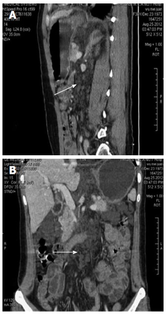
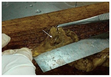
Lymphangioma is an uncommon benign tumor that develops in the lymphatic system. Abdominal lymphangiomatosis is extremely rare in adult patients, and the clinical symptoms of this condition are complicated and atypical. We report a case of abdominal lymphangiomatosis in a 38-year-old female who presented with intestinal bleeding and protein-losing enteropathy, as well as lesions in the lung and bones. A computed tomography scan revealed multiple small cystic lesions without enhancement. Histological examination revealed microscopic cysts were submucosal, with walls composed of thin fibrous tissue, and D2-40 stained highlight the lining of the lymphatic channels by immunohistochemical method. We make a comparison with the cases reported before, and also discuss the diagnose of diffuse pulmonary lymphangiomatosis and Gorham's disease.
Keywords: Abdominal lymphangiomatosis; Diffuse pulmonary lymphangiomatosis; Gastrointestinal bleeding; Gorham’s disease; Lymphangioma.
Figures


Similar articles
-
Management of disseminated intra-abdominal lymphangiomatosis with protein-losing enteropathy and intestinal bleeding.South Med J. 1995 Nov;88(11):1156-8. doi: 10.1097/00007611-199511000-00016. South Med J. 1995. PMID: 7481991
-
Diffuse pulmonary lymphangiomatosis: a case report with literature review.Chin Med J (Engl). 2011 Mar;124(5):797-800. Chin Med J (Engl). 2011. PMID: 21518582 Review.
-
A case of intra-abdominal multiple lymphangiomas in an adult in whom the immunological evaluation supported the diagnosis.Eur J Gastroenterol Hepatol. 1999 Mar;11(3):347-51. doi: 10.1097/00042737-199903000-00022. Eur J Gastroenterol Hepatol. 1999. PMID: 10333211
-
Serious complications of pulmonary biopsy in a boy with chylopericardium and suspected pulmonary lymphangiomatosis.BMJ Case Rep. 2010 May 6;2010:bcr08.2009.2206. doi: 10.1136/bcr.08.2009.2206. BMJ Case Rep. 2010. PMID: 22736725 Free PMC article.
-
Disseminated cystic lymphangiomatosis presenting with acute abdomen: report of a case and review of the literature.Hepatogastroenterology. 1999 Jan-Feb;46(25):196-8. Hepatogastroenterology. 1999. PMID: 10228791 Review.
Cited by
-
Emergency presentation of cystic lymphangioma of the colon: A case report and literature review.Int J Surg Case Rep. 2016;24:162-5. doi: 10.1016/j.ijscr.2016.05.045. Epub 2016 May 27. Int J Surg Case Rep. 2016. PMID: 27266826 Free PMC article.
-
Lymphangiomatosis of the ileum with perforation: A case report and review of the literature.Ann Med Surg (Lond). 2019 Mar 29;41:6-10. doi: 10.1016/j.amsu.2019.03.010. eCollection 2019 May. Ann Med Surg (Lond). 2019. PMID: 30992989 Free PMC article.
-
Lymphangiomatosis associated with protein losing enteropathy: A case report.World J Clin Cases. 2021 May 26;9(15):3758-3764. doi: 10.12998/wjcc.v9.i15.3758. World J Clin Cases. 2021. PMID: 34046480 Free PMC article.
-
The molecular mechanism of Gorham syndrome: an update.Front Immunol. 2023 May 5;14:1165091. doi: 10.3389/fimmu.2023.1165091. eCollection 2023. Front Immunol. 2023. PMID: 37215116 Free PMC article. Review.
-
Systemic generalised lymphangiomatosis: unknown aetiology and a challenge to treat.BMJ Case Rep. 2021 Jan 18;14(1):e237331. doi: 10.1136/bcr-2020-237331. BMJ Case Rep. 2021. PMID: 33462010 Free PMC article.
References
-
- Marom EM, Moran CA, Munden RF. Generalized lymphangiomatosis. AJR Am J Roentgenol. 2004;182:1068. - PubMed
-
- Blei F. Lymphangiomatosis: clinical overview. Lymphat Res Biol. 2011;9:185–190. - PubMed
-
- Amadori G, Micciolo R, Poletti A. A case of intra-abdominal multiple lymphangiomas in an adult in whom the immunological evaluation supported the diagnosis. Eur J Gastroenterol Hepatol. 1999;11:347–351. - PubMed
Publication types
MeSH terms
Supplementary concepts
LinkOut - more resources
Full Text Sources
Other Literature Sources
