. 2014 Jun 24:7:54.
doi: 10.3389/fnmol.2014.00054. eCollection 2014.

LRRK2 kinase activity and biology are not uniformly predicted by its autophosphorylation and cellular phosphorylation site status

Affiliations

LRRK2 kinase activity and biology are not uniformly predicted by its autophosphorylation and cellular phosphorylation site status

April Reynolds et al. Front Mol Neurosci. .

Abstract

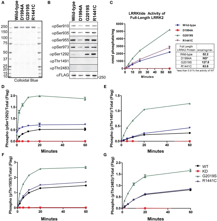
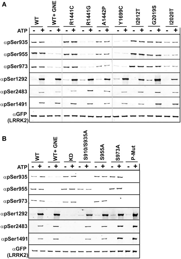
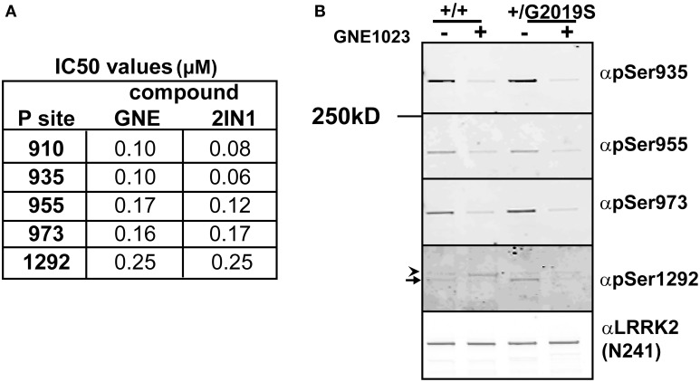
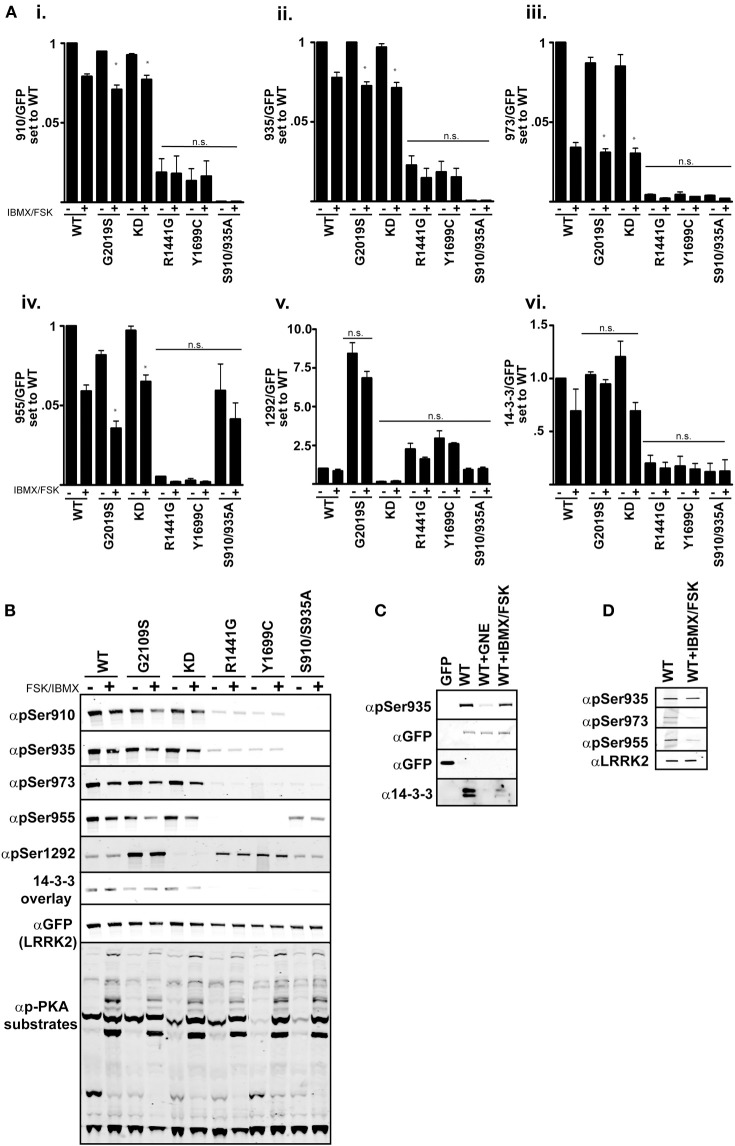
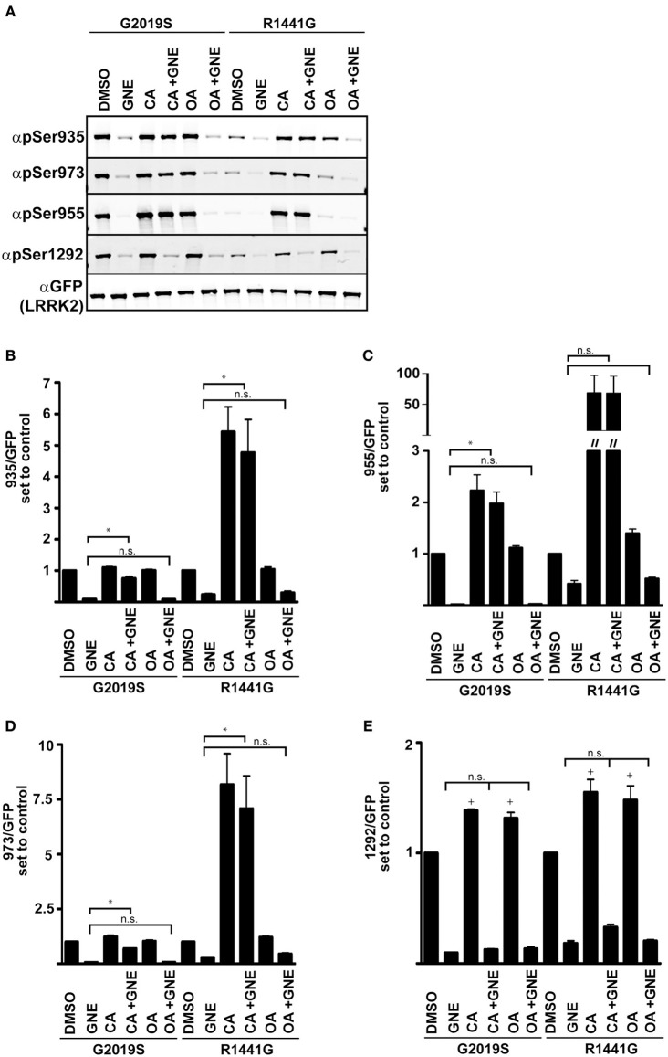
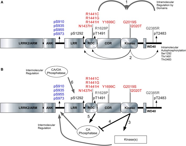
Missense mutations in the Leucine-Rich Repeat protein Kinase 2 (LRRK2) gene are the most common genetic predisposition to develop Parkinson's disease (PD) (Farrer et al., 2005; Skipper et al., 2005; Di Fonzo et al., 2006; Healy et al., 2008; Paisan-Ruiz et al., 2008; Lesage et al., 2010). LRRK2 is a large multi-domain phosphoprotein with a GTPase domain and a serine/threonine protein kinase domain whose activity is implicated in neuronal toxicity; however the precise mechanism is unknown. LRRK2 autophosphorylates on several serine/threonine residues across the enzyme and is found constitutively phosphorylated on Ser910, Ser935, Ser955, and Ser973, which are proposed to be regulated by upstream kinases. Here we investigate the phosphoregulation at these sites by analyzing the effects of disease-associated mutations Arg1441Cys, Arg1441Gly, Ala1442Pro, Tyr1699Cys, Ile2012Thr, Gly2019Ser, and Ile2020Thr. We also studied alanine substitutions of phosphosite serines 910, 935, 955, and 973 and specific LRRK2 inhibition on autophosphorylation of LRRK2 Ser1292, Thr1491, Thr2483 and phosphorylation at the cellular sites. We found that mutants in the Roc-COR domains, including Arg1441Cys, Arg1441His, Ala1442Pro, and Tyr1699Cys, can positively enhance LRRK2 kinase activity, while concomitantly inducing the dephosphorylation of the cellular sites. Mutation of the cellular sites individually did not affect LRRK2 intrinsic kinase activity; however, Ser910/935/955/973Ala mutations trended toward increased kinase activity of LRRK2. Increased cAMP levels did not lead to increased LRRK2 cellular site phosphorylation, 14-3-3 binding or kinase activity. In cells, inhibition of LRRK2 kinase activity leads to dephosphorylation of Ser1292 by Calyculin A and Okadaic acid sensitive phosphatases, while the cellular sites are dephosphorylated by Calyculin A sensitive phosphatases. These findings indicate that comparative analysis of both Ser1292 and Ser910/935/955/973 phosphorylation sites will provide important and distinct measures of LRRK2 kinase and biological activity in vitro and in vivo.

Keywords: GTPase; LRRK2; Parkinson’s disease; kinase; kinase inhibitor; phosphorylation.


Figures

Figure 1
Figure 1
Detection of autophosphorylation and cellular phosphorylation sites on recombinant LRRK2. The indicated full-length recombinant LRRK2 proteins were resolved by SDS-PAGE and stained with either (A) Colloidal Blue to quantify relative protein levels or (B) the indicated phospho-specific antibodies. (C) 14 nM of recombinant wild type LRRK2 (WT) or mutant LRRK2 [Asp1994Ala, Gly2019Ser, Arg1441Cys] in vitro kinase activity was measured against LRRKtide. Incorporated 32P was assessed by Cerenkov counting and results are representative of three independent experiments. Error bars represent mean ± SD. (D–G) 140 nM recombinant LRRK2 was incubated with Mg2+/ATP over a timecourse of 60 min where reaction aliquots were stopped by 1:1 (vol:vol) dilution in 200 mM EDTA and analyzed by dot-blot immunoblot with rabbit monoclonal anti-phospho-Ser1292 (αpS1292), anti-phospho-Thr2483 (αpThr2483), anti-phospho-Thr1491 (αpThr1491) and rabbit polyclonal anti-phospho-Thr1503 antibodies. Results are representative of three independent experiments performed in duplicate and blotted in duplicate. Error bars represent mean ± SD.
Figure 2
Figure 2
Comparison of autophosphorylation and cellular phosphorylation sites in PD-associated mutants. Stable-inducible HEK 293 T-REx cell lines harboring the indicated forms of LRRK2 were incubated for 48 h with 1 μg/ml doxycycline to induce expression. The GFP-LRRK2 WT line was treated with 1 μM GNE1023 for 90 min, before immunoprecipitation-kinase assay using GFP beads, in the presence (+) or absence (−) of ATP. LiCor immunoblot analysis of (A) WT and PD associated mutants of LRRK2 and (B) WT and cellular phosphorylation mutants. Reaction products were probed with rabbit monoclonal anti-phospho-Ser935 (αpS935), anti-phospho-Ser955 (αpS955), anti-phospho-Ser973 (αp973), anti-phospho-Ser1292 (αpS1292), anti-phospho-Thr2483 (αpThr2483), anti-phospho-Thr1491 (αpThr1491). Blots were probed with anti-GFP (αGFP) for total protein control. Phosphorylation levels are quantitated in Supplemental Figure 2.
Figure 3
Figure 3
Comparative analysis of inhibitor induced dephosphorylation for Ser1292 and cellular phosphorylation sites after kinase inhibition. (A) HEK 293 T-REx cells stably expressing GFP-LRRK2-Gly2019Ser were treated with DMSO or increasing concentrations of GNE1023 or LRRK2-IN1 for 90 min. Cell lysates were co-immunoblotted for GFP and anti-phospho-Ser935 (αpS935), anti-phospho-Ser955 (αpS955), anti-phospho-Ser973 (αp973), anti-phospho-Ser1292 (αpS1292). IC50 values were calculated using non-linear regression analysis using GraphPad Prism 6.0. (B) Detection of endogenous pSer1292 in human lymphocytes. Cultures of control donor and +/Gly2019Ser donor lymphoblasts were treated with GNE1023 for 90 min. Endogenous LRRK2 was immunoprecipitated and subjected to immunoblot analysis with the indicated phospho-antibodies. Blots were co-probed with N241 monoclonal antibody to detect total endogenous protein. Arrowhead indicates non-specific band and arrow indicates LRRK2.
Figure 4
Figure 4
cAMP stimulation does not increase 14-3-3 binding in HEK 293 cells. (Ai–vi) Quantification of the phosphorylation levels at the cellular sites (935, 955, 973), autophosphorylation site 1292, and 14-3-3 binding in IBMX/FSK treatment relative to the DMSO treated control from (B). (B) Representative blots of cellular site phosphorylation levels and autophosphorylation at Ser1292 in the indicated GFP-LRRK2 mutants after 30 min treatment with 50 μM FSK/100 μM IBMX. Stimulation of PKA activity is verified by immunoblotting cell lysates with phospho-PKA substrate antibody (Cell Signaling). Results are representative of 5 independent experiments for WT, Gly2019Ser, and KD mutants, and 2 independent experiments for the Arg1441Gly, Tyr1699Cys, and S910/935A mutants. The ratio of phosphorylation over total GFP signal was normalized to WT DMSO, error bars represent s.e.m. Statistical significance was assessed using the One-Way ANOVA test combined with Tukey’s correction. *p ≤ 0.05. Quantification of WT samples treated with DMSO and IBMX/FSK are included in the graphs for reference, but were excluded in the statistical analysis. (C) LRRK2 interaction with 14-3-3 in cells is diminished by IBMX/FSK treatment. GFP or GFP-LRRK2 WT expressing HEK 293 T-REx cells were treated with GNE1023 for 90 min or 50 μM FSK/100 μM IBMX for 30 min, or in combination, then subjected to GFP Trap immunoprecipitation. Immunoprecipitates were immunoblotted for 14-3-3 co-immunoprecipitation. (D) Lung epithelial alveolar A549 cells were treated with 50 μM FSK/100 μM IBMX for 30 min and endogenous LRRK2 was immunoprecipitated and analyzed by immunoblot with anti-LRRK2 (N241), anti-phospho-Ser935, anti-phospho-955, anti-phospho-973 antibodies.
Figure 5
Figure 5
Serine 1292 is regulated by Calyculin A and Okadaic acid sensitive phosphatases. Stable-inducible HEK 293 T-REx cell lines overexpressing Gly2019Ser or Arg1441Gly LRRK2 mutation were treated with Okadaic acid or Calyculin A alone, or combined with GNE1023. (A) Representative blots of phosphorylation levels in Gly2019Ser and Arg1441Gly LRRK2 mutation line. (B–E) Quantification of anti-phospho-Ser935, anti-phospho-955, anti-phospho-973, and anti-phospho-1292 phosphorylation levels relative to the DMSO treated control of each mutant. The immunoblots shown are representative of three independent experiments. Statistical significance of Ser1292 stimulation with CA and OA was assessed using a one sample t-test set to the hypothetical value of 1, +p ≤ 0.05. An ordinary One-Way ANOVA test combined with Dunnett’s correction for multiple testing was used to assess the ability of CA and OA to overcome GNE inhibition *p ≤ 0.05 on connecting bars, n.s., not significant. DMSO control treated samples were not included in the ANOVA test and treatments were compared to GNE (alone) treated samples.
Figure 6
Figure 6
Model of intramolecular and intermolecular LRRK2 regulation. The LRRK2 structure with PD associated mutants highlighted in red (pathogenic) and risk factors in gray. Cellular phosphorylation sites are highlighted in blue and autophosphorylation sites are in black. (A1) The intramolecular regulation of the PD associated mutants in the Roc-COR domain and the carboxy terminus on LRRK2 kinase activity. (A2) Differential effects of the PD mutations can be detected through the autophosphorylation sites Ser1292, Thr1491, and Ser2483. (B3,B4) Kinases and phosphatases contribute to the intermolecular regulation of LRRK2 and their effects can be demonstrated through cellular phosphorylation sites and autophosphorylation sites. (B5) Roc-COR mutations enhance the dephosphorylation of LRRK2 at the cellular sites through increased interaction with phosphatases. Ser1292 is dephosphorylated by Okadaic acid and Calyculin A sensitive phosphatases, (B6) Mutations within the Roc-COR allow for increased interactions with phosphatases and this decreases the cellular phosphorylation levels.

Similar articles

Cited by

References

    1. Adams J. R., Van Netten H., Schulzer M., Mak E., McKenzie J., Strongosky A., et al. (2005). PET in LRRK2 mutations: comparison to sporadic Parkinson’s disease and evidence for presymptomatic compensation. Brain 128, 2777–2785 10.1093/brain/awh607 - DOI - PubMed
    1. Alegre-Abarrategui J., Christian H., Lufino M. M., Mutihac R., Venda L. L., Ansorge O., et al. (2009). LRRK2 regulates autophagic activity and localizes to specific membrane microdomains in a novel human genomic reporter cellular model. Hum. Mol. Genet. 18, 4022–4034 10.1093/hmg/ddp346 - DOI - PMC - PubMed
    1. Baptista M. A., Dave K. D., Frasier M. A., Sherer T. B., Greeley M., Beck M. J., et al. (2013). Loss of leucine-rich repeat kinase 2 (LRRK2) in rats leads to progressive abnormal phenotypes in peripheral organs. PLoS ONE 8:e80705 10.1371/journal.pone.0080705 - DOI - PMC - PubMed
    1. Biosa A., Trancikova A., Civiero L., Glauser L., Bubacco L., Greggio E., et al. (2013). GTPase activity regulates kinase activity and cellular phenotypes of Parkinson’s disease-associated LRRK2. Hum. Mol. Genet. 22, 1140–1156 10.1093/hmg/dds522 - DOI - PubMed
    1. Biskup S., West A. B. (2009). Zeroing in on LRRK2-linked pathogenic mechanisms in Parkinson’s disease. Biochim. Biophys. Acta 1792, 625–633 10.1016/j.bbadis.2008.09.015 - DOI - PMC - PubMed