. 2014 Jun 25:8:53.
doi: 10.3389/fnana.2014.00053. eCollection 2014.

Three axonal projection routes of individual pyramidal cells in the ventral CA1 hippocampus

Affiliations

Three axonal projection routes of individual pyramidal cells in the ventral CA1 hippocampus

Antónia Arszovszki et al. Front Neuroanat. .

Abstract

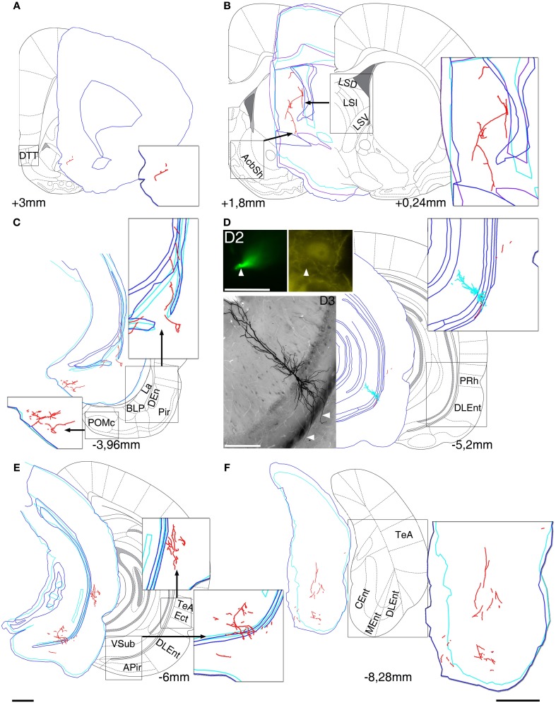
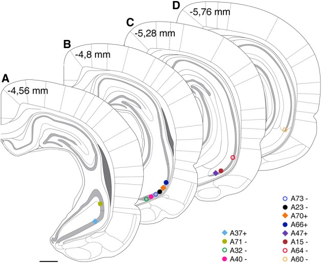
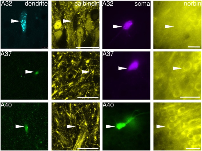
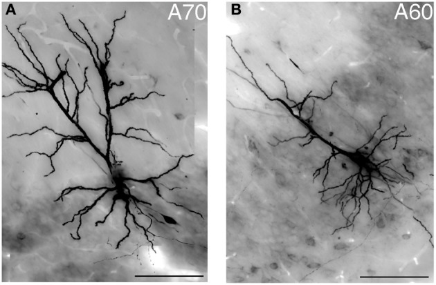
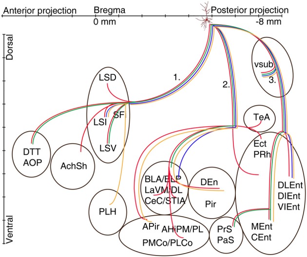
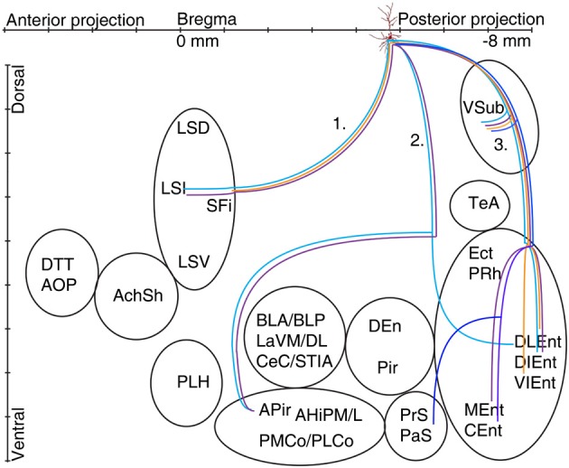
Pyramidal cells of the ventral hippocampal CA1 area have numerous and diverse distant projections to other brain regions including the temporal and parietal association areas, visual, auditory, olfactory, somatosensory, gustatory, and visceral areas, and inputs to the amygdalar and prefrontal-orbital-agranular insular region. In addition, their differential expression of proteins like calbindin provides further indications for cellular diversity. This raises the possibility that the pyramidal cells may form subpopulations participating in different brain circuitries. To address this hypothesis we applied the juxtacellular labeling technique to fill individual pyramidal cells in the ventral hippocampus with neurobiotin in urethane anesthetized rats. For each labeled pyramidal cell we determined soma location, dendritic arborizations and selective expression of calbindin and norbin. Reconstruction and mapping of long-range axonal projections were made with the Neurolucida system. We found three major routes of ventral CA1 pyramidal cell projections. The classical pathway run caudo-ventrally across and innervating the subiculum, further to the parahippocampal regions and then to the deep and superficial layers of entorhinal cortex. The other two pathways avoided subiculum by branching from the main axon close to the soma and either traveled antero- and caudo-ventrally to amygdaloid complex, amygdalopiriform-transition area and parahippocampal regions or run antero-dorsally through the fimbria-fornix to the septum, hypothalamus, ventral striatum and olfactory regions. We found that most pyramidal cells investigated used all three major routes to send projecting axons to other brain areas. Our results suggest that the information flow through the ventral hippocampus is distributed by wide axonal projections from the CA1 area.

Keywords: apical dendrite; axonal projection; calbindin; norbin; pyramidal neuron; ventral CA1.


Figures

Figure 1
Figure 1
Reconstruction of the axonal projections and somato-dendritic position of a labeled pyramidal neuron in VCA1 (A64). Each panel demonstrate a block of several consecutive sections (A/5 section, B/20, C/5, D/2, E/5, and F/5) reconstructed in neurolucida. Each block was correlated with the modified Paxinos atlas along the rostro-caudal axis, showing target areas where local collaterals with boutons (possible terminals) were found. A main axon (thick, myelinated) enters to the septum, and there it give rise to several preterminal axons innervating different subareas (B). More anterior it bifurcates again and simultaneously innervates AcbSh (B) and DTT (A). Different nuclei of amygdalar complex, amygdalopiriform transition area (C) and temporal association cortex TeA (E) are directly innervated. (D) Soma position as reconstructed (red open circle on Figure 2). Cell was calbindin (CB) negative tested on a proximal dendrite (arrowheads, D2). Note that the basal dendrites of the neuron perforate the alveus and enter the deep layer of lateral entorhinal cortex (arrowheads on extended focal image of 2 sections on bottom left). The caudal projection via subiculum emits local collaterals in subiculum (E) and terminates in different parts of the entorhinal cortex (F). Drawings/atlas scale bar: 1 mm; D2/Fluorescent image scale bar 20 μm, D3/Light microscopic image scale bar: 200 μm.
Figure 2
Figure 2
Estimated cell body positions of neurobiotin labeled pyramidal neurons in VCA1 are projected onto coronal sections (A–D) modified from Paxinos atlas. Individually color-coded (see later figures) shapes represents the cells included in this article: Positions of cells are represented by filled circles or filled squares, representing neurons with projection areas shown on Figure 6, or open circles for cells shown in Figure 5. Scale bar: 1 mm.
Figure 3
Figure 3
Fluorescence micrographs showing distinct immunoreactivity of individual VCA1 pyramidal cells for calbindin and norbin. Immunoreactivity for calbindin was tested on apical dendrites of labeled pyramidal cells (confocal images), immunoreactivity for norbin was investigated on labeled somata (Arrowheads) with an epifluorescence microscope. The three cells shown have different immunoreactivity, pyramidal cell A32 was tested as CB negative/norbin negative, cell A37 was double positive and cell A40 was tested negative for CB but positive for norbin. Scale bars: left, 25 μm; right, 20 μm.
Figure 4
Figure 4
Light micrograph showing two distinct geometries of apical dendrites for VCA1 pyramidal cells. (A) Cell A70 has a bifurcated apical dendrite (images were taken by extended focal imaging from 3 consecutive sections with a total thickness of 210 μm), (B) cell A60 has a single apical dendrite (extended focal image from 1 section of 70 μm thickness). Scale bars: 50 μm.
Figure 5
Figure 5
Schematic illustration of the axonal projections and target areas of four CB-negative VCA1 pyramidal cells (A64 red, A60 orange, A32 green, A73 blue same color code as in Figure 2). The main axons of these cells branch in stratum oriens into three different routes: (route 1) via the fimbria/fornix system axons innervate septal nuclei and branches are reaching anterior olfactory regions (A64, A32) or (A60) turn ventrally to innervate the lateral hypothalamus. Axons of route 2 perforate the alveus and project to the amygdalar complex, amygdalohippocampal transition area or the entorhinal cortex. Branches of route 3 cross the ventral subiculum caudally from the soma to different subregions of the entorhinal cortex. (See abbreviation list).
Figure 6
Figure 6
Schematic illustration of the axonal projections and target areas for all labeled CB+ cells (A37 light blue, A47 violet, A70 orange, A66 dark blue). The first route reaches the septum and innervates the intermediate part (A37, A47), the second projection (not observed for cell A70 and A66) arrive to the amygdalopiriform area and cell A37 innervates entorhinal cortex (DLEnt) directly through alveus. In the third direction the cells innervate the subiculum and different parts of the entorhinal cortex.

References

    1. Airaksinen M. S., Eilers J., Garaschuk O., Thoenen H., Konnerth A., Meyer M. (1997). Ataxia and altered dendritic calcium signaling in mice carrying a targeted null mutation of the calbindin D28k gene. Proc. Natl. Acad. Sci. U.S.A. 94, 1488–1493 10.1073/pnas.94.4.1488 - DOI - PMC - PubMed
    1. Amaral D. G., Dolorfo C., Alvarez-Royo P. (1991). Organization of CA1 projections to the subiculum: a PHA-L analysis in the rat. Hippocampus 1, 415–435 10.1002/hipo.450010410 - DOI - PubMed
    1. Amaral D. G., Lavenex P. (2007). Hippocampal neuroanatomy, in The Hippocampus Book, eds Andersen P., Morris R., Amaral D., Bliss T., O'Keefe J. (New York, NY: Oxford University Press; ), 872
    1. Amaral D. G., Witter M. P. (1989). The three-dimensional organization of the hippocampal formation: a review of anatomical data. Neuroscience 31, 571–591 10.1016/0306-4522(89)90424-7 - DOI - PubMed
    1. Andersen P., Bliss T. V., Skrede K. K. (1971). Lamellar organization of hippocampal pathways. Exp. Brain Res. 13, 222–238 10.1007/BF00234087 - DOI - PubMed

LinkOut - more resources