Review
. 2014 Jun 25:5:238.
doi: 10.3389/fphys.2014.00238. eCollection 2014.

The nucleus of the solitary tract and the coordination of respiratory and sympathetic activities

Affiliations
Review

The nucleus of the solitary tract and the coordination of respiratory and sympathetic activities

Daniel B Zoccal et al. Front Physiol. .

Abstract

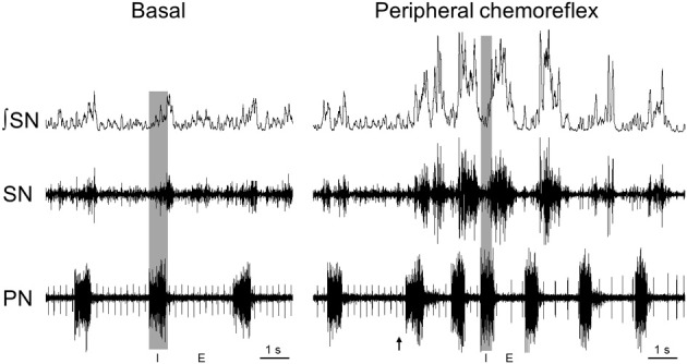
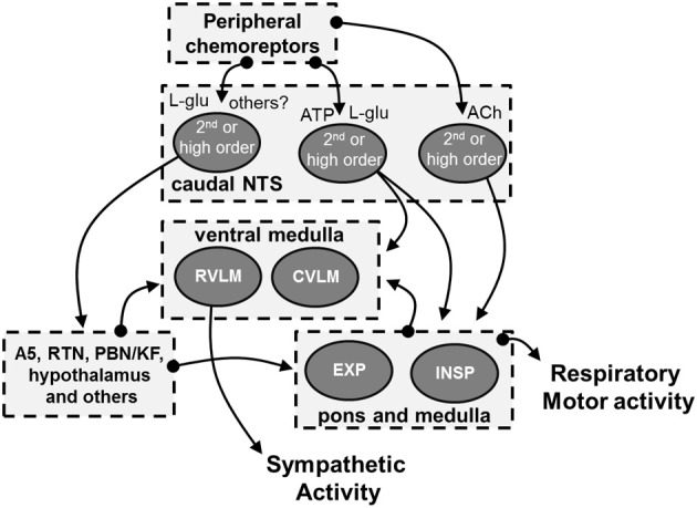
It is well known that breathing introduces rhythmical oscillations in the heart rate and arterial pressure levels. Sympathetic oscillations coupled to the respiratory activity have been suggested as an important homeostatic mechanism optimizing tissue perfusion and blood gas uptake/delivery. This respiratory-sympathetic coupling is strengthened in conditions of blood gas challenges (hypoxia and hypercapnia) as a result of the synchronized activation of brainstem respiratory and sympathetic neurons, culminating with the emergence of entrained cardiovascular and respiratory reflex responses. Studies have proposed that the ventrolateral region of the medulla oblongata is a major site of synaptic interaction between respiratory and sympathetic neurons. However, other brainstem regions also play a relevant role in the patterning of respiratory and sympathetic motor outputs. Recent findings suggest that the neurons of the nucleus of the solitary tract (NTS), in the dorsal medulla, are essential for the processing and coordination of respiratory and sympathetic responses to hypoxia. The NTS is the first synaptic station of the cardiorespiratory afferent inputs, including peripheral chemoreceptors, baroreceptors and pulmonary stretch receptors. The synaptic profile of the NTS neurons receiving the excitatory drive from afferent inputs is complex and involves distinct neurotransmitters, including glutamate, ATP and acetylcholine. In the present review we discuss the role of the NTS circuitry in coordinating sympathetic and respiratory reflex responses. We also analyze the neuroplasticity of NTS neurons and their contribution for the development of cardiorespiratory dysfunctions, as observed in neurogenic hypertension, obstructive sleep apnea and metabolic disorders.

Keywords: NTS; cardiorespiratory coupling; chemoreflex; neurotransmission; respiration; sympathetic activity.


Figures

Figure 1
Figure 1
Respiratory-sympathetic coupling at basal conditions and during peripheral chemoreflex activation. Raw and integrated (⨛) recordings obtained in a decerebrated, arterially-perfused in situ rat preparation (for details, please see Zoccal et al., 2008) showing the coupling of phrenic (PN) and thoracic sympathetic nerve activity (SN) at basal conditions (normocapnia) and during peripheral chemoreflex activation, evoked by intra-arterial administration of potassium cyanide (0.05%, 50 μL; arrow). The shaded gray area delimitates the inspiratory phase (I; coincident with phrenic burst) while the phrenic burst interval correspond to expiratory phase (E).
Figure 2
Figure 2
Schematic drawing showing the possible cellular and neurochemical mechanisms of the cNTS mediating the processing of peripheral chemoreceptors inputs. The stimulation of peripheral chemoreceptors evokes the release of L-glutamate in the cNTS (Mifflin, ; Andresen and Kunze, 1994). Experimental evidence indicates the L-glutamate, in association with ATP, is essential for the processing of sympatho-excitatory response of the peripheral chemoreflex in the cNTS (Machado and Bonagamba, ; Braga et al., 2007). It is suggested that both glutamatergic and purinergic systems interact and activate cNTS neurons that send projections to pre-sympathetic neurons of the RVLM (Accorsi-Mendonca et al., 2009, 2013). L-glutamate is also proposed to mediate the activation of other neural pathways that are important for the sympatho-excitatory component of peripheral chemoreflex, including those to the A5 region, retrotrapezoid nucleus (RTN), parabrachial nucleus/Kölliker-Fuse complex (PBN/KF) and hypothalamus (Koshiya and Guyenet, ; Olivan et al., ; Haibara et al., ; Reddy et al., ; Takakura et al., ; Queiroz et al., ; Song et al., ; Taxini et al., ; King et al., 2012). In addition to L-glutamate and ATP, our recent studies suggest that ACh in the cNTS significantly contributes to the tachypnea and the patterning of sympathetic response of peripheral chemoreflex (Furuya et al., 2014). We hypothesize that ACh activates cNTS neurons that send projections to respiratory neurons of the ventral medulla that, in turn, promotes the stimulation of inspiratory motor activity and the patterning of sympathetic activity. The latter may involve the modulation of the neurons of RVLM and of the caudal ventrolateral medulla (CVLM) (Mandel and Schreihofer, ; Moraes et al., 2012c). However, it remains to be elucidated the sources of ACh in the cNTS as well as the efferent pathways activated by ACh in response to peripheral chemoreflex activation.

Similar articles

Cited by

References

    1. Abdala A. P., McBryde F. D., Marina N., Hendy E. B., Engelman Z. J., Fudim M., et al. (2012). Hypertension is critically dependent on the carotid body input in the spontaneously hypertensive rat. J. Physiol. 590, 4269–4277 10.1113/jphysiol.2012.237800 - DOI - PMC - PubMed
    1. Accorsi-Mendonca D., Bonagamba L. G., Leao R. M., Machado B. H. (2009). Are L-glutamate and ATP cotransmitters of the peripheral chemoreflex in the rat nucleus tractus solitarius? Exp. Physiol. 94, 38–45 10.1113/expphysiol.2008.043653 - DOI - PubMed
    1. Accorsi-Mendonca D., Castania J. A., Bonagamba L. G., Machado B. H., Leao R. M. (2011). Synaptic profile of nucleus tractus solitarius neurons involved with the peripheral chemoreflex pathways. Neuroscience 197, 107–120 10.1016/j.neuroscience.2011.08.054 - DOI - PubMed
    1. Accorsi-Mendonca D., Zoccal D. B., Bonagamba L. G., Machado B. H. (2013). Glial cells modulate the synaptic transmission of NTS neurons sending projections to ventral medulla of Wistar rats. Physiol. Rep. 1:e00080 10.1002/phy2.80 - DOI - PMC - PubMed
    1. Adrian E. D., Bronk D. W., Phillips G. (1932). Discharges in mammalian sympathetic nerves. J. Physiol. 74, 115–133 - PMC - PubMed