

Effects of exogenous pyoverdines on Fe availability and their impacts on Mn(II) oxidation by Pseudomonas putida GB-1
- PMID: 25009534
- PMCID: PMC4070179
- DOI: 10.3389/fmicb.2014.00301
Effects of exogenous pyoverdines on Fe availability and their impacts on Mn(II) oxidation by Pseudomonas putida GB-1
Abstract
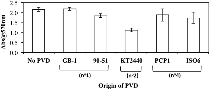
Pseudomonas putida GB-1 is a Mn(II)-oxidizing bacterium that produces pyoverdine-type siderophores (PVDs), which facilitate the uptake of Fe(III) but also influence MnO2 formation. Recently, a non-ribosomal peptide synthetase mutant that does not synthesize PVD was described. Here we identified a gene encoding the PVDGB-1 (PVD produced by strain GB-1) uptake receptor (PputGB1_4082) of strain GB-1 and confirmed its function by in-frame mutagenesis. Growth and other physiological responses of these two mutants and of wild type were compared during cultivation in the presence of three chemically distinct sets of PVDs (siderotypes n°1, n°2, and n°4) derived from various pseudomonads. Under iron-limiting conditions, Fe(III) complexes of various siderotype n°1 PVDs (including PVDGB-1) allowed growth of wild type and the synthetase mutant, but not the receptor mutant, confirming that iron uptake with any tested siderotype n°1 PVD depended on PputGB1_4082. Fe(III) complexes of a siderotype n°2 PVD were not utilized by any strain and strongly induced PVD synthesis. In contrast, Fe(III) complexes of siderotype n°4 PVDs promoted the growth of all three strains and did not induce PVD synthesis by the wild type, implying these complexes were utilized for iron uptake independent of PputGB1_4082. These differing properties of the three PVD types provided a way to differentiate between effects on MnO2 formation that resulted from iron limitation and others that required participation of the PVDGB-1 receptor. Specifically, MnO2 production was inhibited by siderotype n°1 but not n°4 PVDs indicating PVD synthesis or PputGB1_4082 involvement rather than iron-limitation caused the inhibition. In contrast, iron limitation was sufficient to explain the inhibition of Mn(II) oxidation by siderotype n°2 PVDs. Collectively, our results provide insight into how competition for iron via siderophores influences growth, iron nutrition and MnO2 formation in more complex environmental systems.
Keywords: Fe availability; MnO2; biofilm; iron limitation; iron requirement; pyoverdine; pyoverdine receptor; siderotyping.
Figures
Similar articles
-
Pyoverdine synthesis by the Mn(II)-oxidizing bacterium Pseudomonas putida GB-1.Front Microbiol. 2014 May 7;5:202. doi: 10.3389/fmicb.2014.00202. eCollection 2014. Front Microbiol. 2014. PMID: 24847318 Free PMC article.
-
Pyoverdines Are Essential for the Antibacterial Activity of Pseudomonas chlororaphis YL-1 under Low-Iron Conditions.Appl Environ Microbiol. 2021 Mar 11;87(7):e02840-20. doi: 10.1128/AEM.02840-20. Print 2021 Mar 11. Appl Environ Microbiol. 2021. PMID: 33452032 Free PMC article.
-
Siderotyping of fluorescent pseudomonads: characterization of pyoverdines of Pseudomonas fluorescens and Pseudomonas putida strains from Antarctica.Microbiology (Reading). 1998 Nov;144 ( Pt 11):3119-3126. doi: 10.1099/00221287-144-11-3119. Microbiology (Reading). 1998. PMID: 9846748
-
Metal trafficking via siderophores in Gram-negative bacteria: specificities and characteristics of the pyoverdine pathway.J Inorg Biochem. 2008 May-Jun;102(5-6):1159-69. doi: 10.1016/j.jinorgbio.2007.11.017. Epub 2007 Dec 14. J Inorg Biochem. 2008. PMID: 18221784 Review.
-
Diversity of siderophore-mediated iron uptake systems in fluorescent pseudomonads: not only pyoverdines.Environ Microbiol. 2002 Dec;4(12):787-98. doi: 10.1046/j.1462-2920.2002.00369.x. Environ Microbiol. 2002. PMID: 12534462 Review.
Cited by
-
bifA Regulates Biofilm Development of Pseudomonas putida MnB1 as a Primary Response to H2O2 and Mn2.Front Microbiol. 2018 Jul 10;9:1490. doi: 10.3389/fmicb.2018.01490. eCollection 2018. Front Microbiol. 2018. PMID: 30042743 Free PMC article.
-
The Role of Fur in the Transcriptional and Iron Homeostatic Response of Enterococcus faecalis.Front Microbiol. 2018 Jul 17;9:1580. doi: 10.3389/fmicb.2018.01580. eCollection 2018. Front Microbiol. 2018. PMID: 30065712 Free PMC article.
References
Grants and funding
LinkOut - more resources
Full Text Sources
Other Literature Sources