

RCAN1 regulates mitochondrial function and increases susceptibility to oxidative stress in mammalian cells
- PMID: 25009690
- PMCID: PMC4070399
- DOI: 10.1155/2014/520316
RCAN1 regulates mitochondrial function and increases susceptibility to oxidative stress in mammalian cells
Abstract
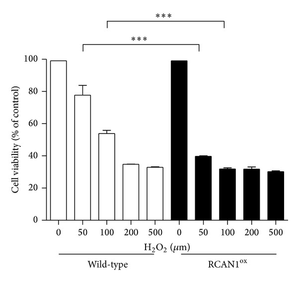
Mitochondria are the primary site of cellular energy generation and reactive oxygen species (ROS) accumulation. Elevated ROS levels are detrimental to normal cell function and have been linked to the pathogenesis of neurodegenerative disorders such as Down's syndrome (DS) and Alzheimer's disease (AD). RCAN1 is abundantly expressed in the brain and overexpressed in brain of DS and AD patients. Data from nonmammalian species indicates that increased RCAN1 expression results in altered mitochondrial function and that RCAN1 may itself regulate neuronal ROS production. In this study, we have utilized mice overexpressing RCAN1 (RCAN1(ox)) and demonstrate an increased susceptibility of neurons from these mice to oxidative stress. Mitochondria from these mice are more numerous and smaller, indicative of mitochondrial dysfunction, and mitochondrial membrane potential is altered under conditions of oxidative stress. We also generated a PC12 cell line overexpressing RCAN1 (PC12(RCAN1)). Similar to RCAN1(ox) neurons, PC12(RCAN1) cells have an increased susceptibility to oxidative stress and produce more mitochondrial ROS. This study demonstrates that increasing RCAN1 expression alters mitochondrial function and increases the susceptibility of neurons to oxidative stress in mammalian cells. These findings further contribute to our understanding of RCAN1 and its potential role in the pathogenesis of neurodegenerative disorders such as AD and DS.
Figures


Similar articles
-
Lycopene inhibits regulator of calcineurin 1-mediated apoptosis by reducing oxidative stress and down-regulating Nucling in neuronal cells.Mol Nutr Food Res. 2017 May;61(5). doi: 10.1002/mnfr.201600530. Epub 2017 Feb 6. Mol Nutr Food Res. 2017. PMID: 27928873
-
RCAN1 (DSCR1) increases neuronal susceptibility to oxidative stress: a potential pathogenic process in neurodegeneration.Hum Mol Genet. 2007 May 1;16(9):1039-50. doi: 10.1093/hmg/ddm049. Epub 2007 Mar 6. Hum Mol Genet. 2007. PMID: 17341486
-
RCAN1 overexpression promotes age-dependent mitochondrial dysregulation related to neurodegeneration in Alzheimer's disease.Acta Neuropathol. 2015 Dec;130(6):829-43. doi: 10.1007/s00401-015-1499-8. Epub 2015 Oct 24. Acta Neuropathol. 2015. PMID: 26497675 Free PMC article.
-
Chronic high levels of the RCAN1-1 protein may promote neurodegeneration and Alzheimer disease.Free Radic Biol Med. 2013 Sep;62:47-51. doi: 10.1016/j.freeradbiomed.2013.01.016. Epub 2013 Jan 29. Free Radic Biol Med. 2013. PMID: 23369757 Free PMC article. Review.
-
The neuronal and endocrine roles of RCAN1 in health and disease.Clin Exp Pharmacol Physiol. 2018 Apr;45(4):377-383. doi: 10.1111/1440-1681.12884. Epub 2017 Nov 29. Clin Exp Pharmacol Physiol. 2018. PMID: 29094385 Review.
Cited by
-
Regulator of calcineurin 1 deletion attenuates mitochondrial dysfunction and apoptosis in acute kidney injury through JNK/Mff signaling pathway.Cell Death Dis. 2022 Sep 7;13(9):774. doi: 10.1038/s41419-022-05220-x. Cell Death Dis. 2022. PMID: 36071051 Free PMC article.
-
Changes in miroRNA-103 expression in wound margin tissue are related to wound healing of diabetes foot ulcers.Int Wound J. 2023 Feb;20(2):467-483. doi: 10.1111/iwj.13895. Epub 2022 Jul 15. Int Wound J. 2023. PMID: 35837786 Free PMC article.
-
A Syntenic Cross Species Aneuploidy Genetic Screen Links RCAN1 Expression to β-Cell Mitochondrial Dysfunction in Type 2 Diabetes.PLoS Genet. 2016 May 19;12(5):e1006033. doi: 10.1371/journal.pgen.1006033. eCollection 2016 May. PLoS Genet. 2016. PMID: 27195491 Free PMC article.
-
Abundance of Synaptic Vesicle-Related Proteins in Alpha-Synuclein-Containing Protein Inclusions Suggests a Targeted Formation Mechanism.Neurotox Res. 2019 May;35(4):883-897. doi: 10.1007/s12640-019-00014-0. Epub 2019 Feb 22. Neurotox Res. 2019. PMID: 30796693
-
Down Syndrome Critical Region 1 Gene, Rcan1, Helps Maintain a More Fused Mitochondrial Network.Circ Res. 2018 Mar 16;122(6):e20-e33. doi: 10.1161/CIRCRESAHA.117.311522. Epub 2018 Jan 23. Circ Res. 2018. PMID: 29362227 Free PMC article.
References
-
- Fuentes J-J, Pritchard MA, Planas AM, Bosch A, Ferrer I, Estivill X. A new human gene from the down syndrome critical region encodes a proline-rich protein highly expressed in fetal brain and heart. Human Molecular Genetics. 1995;4(10):1935–1944. - PubMed
-
- Ermak G, Morgan TE, Davies KJA. Chronic overexpression of the calcineurin inhibitory gene DSCR1 (Adapt78) is associated with Alzheimer’s disease. The Journal of Biological Chemistry. 2001;276(42):38787–38794. - PubMed
-
- Peiris H, Raghupathi R, Jessup CF, et al. Increased expression of the glucose-responsive gene, RCAN1, causes hypoinsulinemia, β-cell dysfunction, and diabetes. Endocrinology. 2012;153(11):5212–5221. - PubMed
-
- Fuentes JJ, Genescà L, Kingsbury TJ, et al. DSCR1, overexpressed in Down syndrome, is an inhibitor of calcineurin-mediated signaling pathways. Human Molecular Genetics. 2000;9(11):1681–1690. - PubMed
-
- Yang J, Rothermel B, Vega RB, et al. Independent signals control expression of the calcineurin inhibitory proteins MCIP1 and MCIP2 in striated muscles. Circulation Research. 2000;87(12):E61–E68. - PubMed
Publication types
MeSH terms
Substances
LinkOut - more resources
Full Text Sources
Other Literature Sources
