. 2014 May;22(3):184-92.
doi: 10.4062/biomolther.2014.026.

β-lapachone-Induced Apoptosis of Human Gastric Carcinoma AGS Cells Is Caspase-Dependent and Regulated by the PI3K/Akt Pathway

Affiliations

β-lapachone-Induced Apoptosis of Human Gastric Carcinoma AGS Cells Is Caspase-Dependent and Regulated by the PI3K/Akt Pathway

Hai Yang Yu et al. Biomol Ther (Seoul). 2014 May.

Abstract

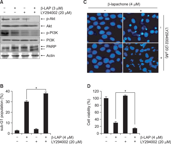
β-lapachone is a naturally occurring quinone that selectively induces apoptotic cell death in a variety of human cancer cells in vitro and in vivo; however, its mechanism of action needs to be further elaborated. In this study, we investigated the effects of β-lapachone on the induction of apoptosis in human gastric carcinoma AGS cells. β-lapachone significantly inhibited cellular proliferation, and some typical apoptotic characteristics such as chromatin condensation and an increase in the population of sub-G1 hypodiploid cells were observed in β-lapachone-treated AGS cells. Treatment with β-lapachone caused mitochondrial transmembrane potential dissipation, stimulated the mitochondria-mediated intrinsic apoptotic pathway, as indicated by caspase-9 activation, cytochrome c release, Bcl-2 downregulation and Bax upregulation, as well as death receptor-mediated extrinsic apoptotic pathway, as indicated by activation of caspase-8 and truncation of Bid. This process was accompanied by activation of caspase-3 and concomitant with cleavage of poly(ADP-ribose) polymerase. The general caspase inhibitor, z-VAD-fmk, significantly abolished β-lapachone-induced cell death and inhibited growth. Further analysis demonstrated that the induction of apoptosis by β-lapachone was accompanied by inactivation of the phosphatidylinositol 3-kinase (PI3K)/Akt signaling pathway. The PI3K inhibitor LY29004 significantly increased β-lapachone-induced apoptosis and growth inhibition. Taken together, these findings indicate that the apoptotic activity of β-lapachone is probably regulated by a caspase-dependent cascade through activation of both intrinsic and extrinsic signaling pathways, and that inhibition of the PI3K/Akt signaling may contribute to β-lapachone-mediated AGS cell growth inhibition and apoptosis induction.

Keywords: Apoptosis; Caspase; PI3K/Akt; β-lapachone.


Figures

Fig. 1.
Fig. 1.
β-lapachone inhibits cell viability in AGS cells. Cells were seeded at 2×10⁵ cell per well onto 96-well culture plates overnight and then treated with the indicated concentrations of β-lapachone for 24 h. Cell viability was determined by the MTT assay. Data are mean ± SD of three independent experiments (*p<0.05 vs. untreated control).
Fig. 2.
Fig. 2.
β-lapachone induces apoptosis in AGS cells. (A) The cells were treated with the indicated concentrations of β-lapachone for 24 h, sampled, fixed, and stained with DAPI solution. The stained nuclei were observed under a fluorescent microscope (original magnification, 400×). (B) To quantify the degree of apoptosis induced by β-lapachone, cells grown under the same conditions as (A) were evaluated by a flow cytometry for sub-G1 DNA content, which represents the cells undergoing apoptotic DNA degradation. Data are mean ± SD of three independent experiments (*p<0.05 vs. untreated control).
Fig. 3.
Fig. 3.
Activation of caspases and degradation of PARP by β-lapachone in AGS cells. (A) Cells were treated with the indicated concentrations of β-lapachone for 24 h. The cells were lysed, and cellular proteins were separated by SDS-polyacrylamide gel electrophoresis and transferred to nitrocellulose membranes. The proteins were visualized using the indicated antibodies and an ECL detection system. Proteolytic cleavage of PARP is indicated by the arrow. Actin was used as the internal control. (B) After a 24 h incubation with the indicated concentrations of β-lapachone, the cells were lysed, and aliquots were assayed for in vitro caspase-3, -8, and -9 activity using Ac-DEVD-pNA, Ac-IETD-pNA, and Ac-LEHD-pNA as substrates, respectively, at 37°C. After incubation of 1 h, the amount of pNA released was measured at 405 nm using an ELISA microplate reader. Each point represents the mean ± SD of three independent experiments (*p<0.05 vs. untreated control).
Fig. 4.
Fig. 4.
Caspase-mediated apoptosis induced by β-lapachone in AGS cells. (A) Cells were incubated with 4 μM β-lapachone for 24 h after 1 h pretreatment with the pan-caspase inhibitor, z-VAD-fmk (50 μM). Equal amounts of cell lysates isolated from cells were subjected to SDS-polyacrylamide gel electrophoresis and analyzed by Western blot for caspase-3 and PARP. (B) Cells grown under the same conditions as (A) were fixed and stained with DAPI solution. Stained nuclei were observed under a fluorescent microscope. (C) Cells were evaluated by flow cytometry for sub-G1 DNA content, suggestive of apoptotic cell death. (D) Cell viability was determined by the MTT assay. Results are mean ± SD of three independent experiments (*p<0.05 vs. β-lapachone treated cells).
Fig. 5.
Fig. 5.
Effect of β-lapachone on levels of MMP, Bcl-2 family proteins, and cytochrome c in AGS cells. (A) Cells were treated with indicated concentrations of β-lapachone for 24 h. They were collected and incubated with 10 μM JC-1 for 15 min at 37°C in the dark. The cells were washed once with PBS and analyzed by a DNA flow cytometer. Results are presented as the mean of two independent experiments. (B) Equal amounts of cell lysates was resolved by SDS-polyacrylamide gel electrophoresis, transferred to membranes, and probed with specific antibodies. The anti-actin antibody was a protein loading control. (C) Cytosolic and mitochondrial proteins were extracted from cells grown under the same conditions and analyzed by Western blotting using anti-cytochrome c antibody. Actin and cytochrome oxidase subunit IV (COX IV) were used as internal controls for the cytosolic and mitochondrial fractions, respectively.
Fig. 6.
Fig. 6.
Effect of β-lapachone on the levels of phosphorylation of PI3K and Akt in AGS cells. The cells were incubated with the indicated concentrations of β-lapachone for 24 h. The cells were lysed, and the cellular proteins were separated by SDS-polyacrylamide gel electrophoresis and transferred to nitrocellulose membranes. The membranes were probed with specific antibodies to p-Akt (Ser 473), total Akt, p-PI3K (p85 Tyr 458/p55 Tyr 199), and total PI3K. The proteins were visualized using an ECL detection system. Actin was used as the internal control.
Fig. 7.
Fig. 7.
The role of PI3K/Akt signaling in β-lapachone-induced apoptosis in AGS cells. (A) Cells were stimulated with 3 μM β-lapachone for 24 h after pretreatment with 20 μM LY290042 for 1 h. Equal amounts of cell lysates were resolved by SDS-polyacrylamide gel electrophoresis, transferred to membranes, and probed with the indicated antibodies. Anti-actin antibody was a protein loading control. (B and D) The percentage of the sub-G1 population (B) and cell viability (D) were assessed by flow cytometry and the MTT assay, respectively. Results are mean ± SD of three independent experiments (*p<0.05 vs. β-lapachone treated cells). (C) The cells were fixed and stained with DAPI solution. Stained nuclei were observed under a fluorescent microscope.

Similar articles

Cited by

References

    1. Ang KL, Shi DL, Keong WW, Epstein RJ. Up-regulated Akt signaling adjacent to gastric cancers: implications for screening and chemoprevention. Cancer Lett. 2005;225:53–59. - PubMed
    1. Bey EA, Bentle MS, Reinicke KE, Dong Y, Yang CR, Girard L, Minna JD, Bornmann WG, Gao J, Boothman DA. An NQO1- and PARP-1-mediated cell death pathway induced in non-small-cell lung cancer cells by β-lapachone. Proc Natl Acad Sci USA. 2007;104:11832–11837. - PMC - PubMed
    1. Binutu OA, Adesogan KE, Okogun JI. Antibacterial and antifungal compounds from Kigelia pinnata. Planta Med. 1996;62:352–353. - PubMed
    1. Boothman DA, Trask DK, Pardee AB. Inhibition of potentially lethal DNA damage repair in human tumor cells by β-lapachone, an activator of topoisomerase I. Cancer Res. 1989;49:605–612. - PubMed
    1. Carnero A, Blanco-Aparicio C, Renner O, Link W, Leal JF. The PTEN/PI3K/AKT signalling pathway in cancer, therapeutic implications. Curr. Cancer Drug Targets. 2008;8:187–198. - PubMed

LinkOut - more resources