

Sacro-anterior haemangiopericytoma: a case report
- PMID: 25009757
- PMCID: PMC4069801
- DOI: 10.7497/j.issn.2095-3941.2014.02.010
Sacro-anterior haemangiopericytoma: a case report
Abstract
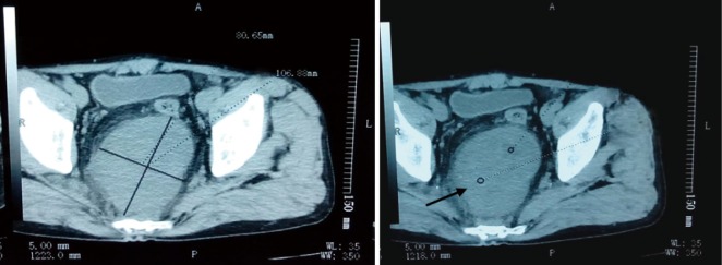
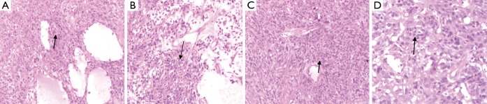
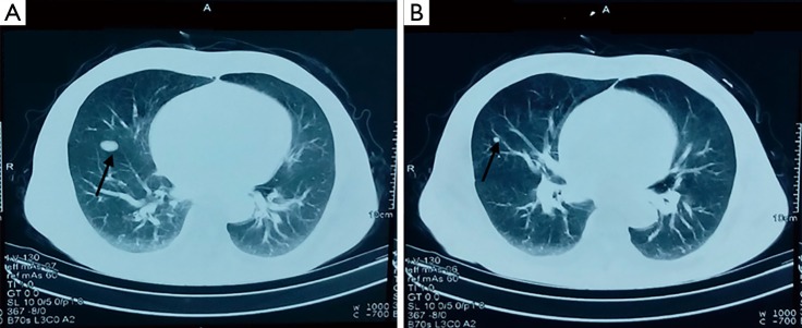
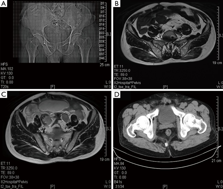
Haemangiopericytoma (HPC) is a rare vascular tumor with borderline malignancy, considerable histological variability, and unpredictable clinical and biological behavior. HPC can present a diagnostic challenge because of its indeterminate clinical, radiological, and pathological features. HPC generally presents in adulthood and is equally frequent in both sexes. HPC can arise in any site in the body as a slowly growing and painless mass. The precise cell type origin of HPC is uncertain. One third of HPCs occur in the head and neck areas. Exceptional cases of hemangioblastoma arising outside the head and neck areas have been reported, but little is known about their clinicopathologic and immunohistochemical features. This study reports on a case of a large sacro-anterior HPC in a 65-year-old male.
Keywords: Sacro-anterior; haemangiopericytoma (HPC); soft tissue tumor.
Conflict of interest statement
No potential conflicts of interest are disclosed.
Figures
Similar articles
-
A rare case of orbital haemangiopericytoma arising in childhood.Orbit. 2013 Dec;32(6):384-6. doi: 10.3109/01676830.2013.815226. Epub 2013 Jul 29. Orbit. 2013. PMID: 23895036
-
Infantile haemangiopericytoma: a rare congenital cervical tumour.J Laryngol Otol. 2014 Feb;128(2):212-5. doi: 10.1017/S0022215113003630. Epub 2014 Feb 10. J Laryngol Otol. 2014. PMID: 24507075
-
Recurrent primary haemangiopericytoma of the bladder: A case report and literature review.Oncol Lett. 2014 Apr;7(4):1144-1146. doi: 10.3892/ol.2014.1862. Epub 2014 Feb 7. Oncol Lett. 2014. PMID: 24944682 Free PMC article.
-
Primary haemangiopericytoma outside muscle cone in fossa orbitalis: a case report and review.Eye Sci. 2012 Dec;27(4):205-9. doi: 10.3969/j.issn.1000-4432.2012.04.011. Eye Sci. 2012. PMID: 23225844 Review.
-
Solitary fibrous tumour and haemangiopericytoma: evolution of a concept.Histopathology. 2006 Jan;48(1):63-74. doi: 10.1111/j.1365-2559.2005.02290.x. Histopathology. 2006. PMID: 16359538 Review.
References
-
- Russell WO, Cohen J, Enzinger F, Hajdu SI, Heise H, Martin RG, et al. A clinical and pathological staging system for soft tissue sarcomas. Cancer 1977;40:1562-1570 - PubMed
-
- Sun Y, Tian JH, Li M, Shi CJ. Two cases of hemangiopericytoma. Journal of Chinese Clinical Oncology 2010,15:284-285
Publication types
LinkOut - more resources
Full Text Sources